A CAD/CAM full arch restoration is a digitally engineered, implant-supported prosthesis designed to replace an entire upper or lower set of teeth. Utilizing advanced 3D scanning and computer-aided milling, this protocol helps ensure precise bite alignment, optimal aesthetics, and the long-lasting durability of monolithic zirconia without relying on traditional, messy impressions.
Clinical Summary:
The transition from analog to digital dentistry has revolutionized complex prosthetic rehabilitations. A CAD/CAM full arch restoration leverages intraoral 3D scanning, precise digital smile design, and automated milling to create highly accurate, biocompatible prostheses. This digital workflow is essential for accurately restoring the vertical dimension of occlusion and supporting temporomandibular joint health. By utilizing monolithic zirconia, clinicians can achieve a margin fit under 50 micrometers, significantly reducing the risk of bacterial microleakage and porcelain chipping. These advanced protocols are often combined with IV conscious sedation and Platelet-Rich Fibrin (PRF) therapy to promote a comfortable, predictable, and biologically sound full mouth rehabilitation.
Key Takeaways:
- Intraoral 3D scanners eliminate the need for uncomfortable silicone putty impressions, providing highly accurate topographical data.
- Restoring the vertical dimension of occlusion is critical for addressing bite collapse and supporting neuromuscular harmony.
- CAD/CAM milling of monolithic zirconia produces restorations with superior flexural strength and precise margin adaptation.
- IV conscious sedation and PRF therapy can enhance patient comfort and support soft tissue and bone healing.
- Digital workflows allow for predictable aesthetic outcomes through comprehensive 3D digital smile design mock-ups.
Medit i700 and iTero 3D: Eliminating Messy Silicone Molds for Full Arch Cases
Advanced intraoral 3D scanners capture highly accurate digital impressions, bypassing the discomfort and dimensional distortion associated with traditional silicone putty molds.
The foundation of any successful CAD/CAM full arch restoration begins with the accurate capture of the patient’s existing oral anatomy. Historically, clinicians relied on polyvinyl siloxane (PVS) or polyether impression materials. While effective, these analog methods presented significant challenges, particularly for patients with a strong gag reflex or severe dental anxiety. Furthermore, traditional impressions are susceptible to dimensional changes due to temperature variations, moisture contamination, and the physical expansion of dental stone during the pouring process.
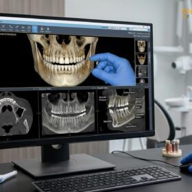
The advent of the intraoral 3D scan has fundamentally shifted this paradigm. Devices such as the Medit i700 and iTero utilize confocal microscopy and structured light technologies to capture thousands of images per second, stitching them together to create a highly accurate, full-color 3D model of the dental arches. This digital intraoral impression is not only infinitely more comfortable for the patient but also provides the dental laboratory with an exact, distortion-free replica of the prepared teeth or implant scan bodies. According to guidelines from the Vietnam Odonto-Stomatology Association (VOSA), the integration of digital scanning is highly recommended to meet modern standards of prosthetic accuracy[6].
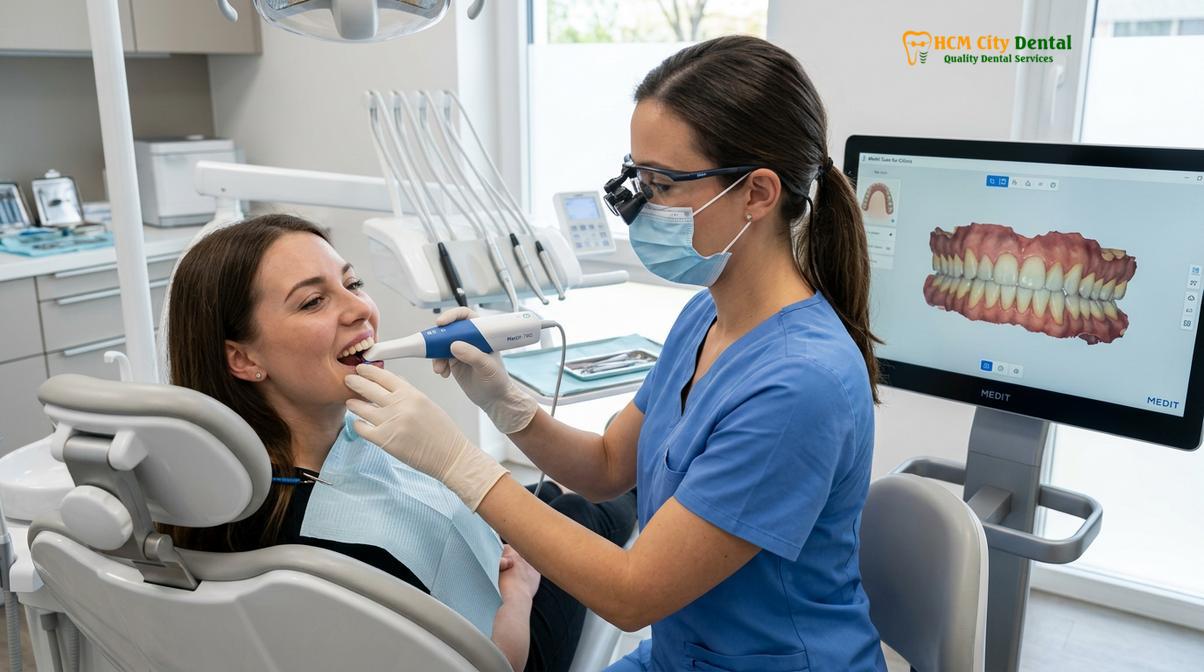
In the context of full mouth rehabilitation, the speed and accuracy of these scanners are paramount. Capturing the precise angulation and depth of multiple dental implants across an entire arch requires sub-millimeter precision. The digital file can be instantly transmitted to the CAD/CAM design software, drastically reducing the turnaround time between the surgical placement of implants and the delivery of the provisional prosthesis. This seamless integration is a cornerstone of modern implantology, ensuring that the final restoration seats passively without inducing undue stress on the integrating titanium fixtures[1].
Digital Workflows: Scanning the Prepared Arches and Bite Relationships
Capturing the exact spatial relationship between the upper and lower jaws is critical for correcting bite collapse and supporting long-term neuromuscular stability.
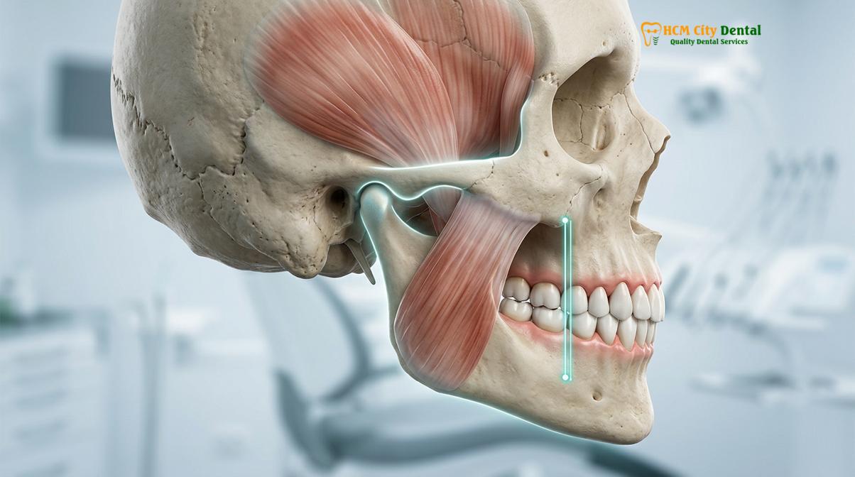
Beyond capturing the physical topography of the teeth and implants, the digital workflow must accurately record the dynamic relationship between the maxilla (upper jaw) and mandible (lower jaw). When a patient suffers from severe tooth loss or severe attrition (wear), they often experience a loss of the vertical dimension of occlusion (VDO). This bite collapse restoration is one of the most complex challenges in prosthodontics, as it directly impacts facial aesthetics, chewing efficiency, and the health of the temporomandibular joint (TMJ).
To address this, clinicians employ principles of neuromuscular dentistry. The goal is to find the physiologic rest jaw position—the point where the masticatory muscles are at their most relaxed state. Techniques such as Transcutaneous Electrical Nerve Stimulation (TENS) therapy may be used to induce jaw muscle relaxation, allowing the clinician to record the bite in its optimal, unforced trajectory. Once this ideal position is determined, a diagnostic splint (an orthotic device) is often fabricated. The patient wears this splint temporarily to verify that the new jaw position is comfortable and functional before any permanent alterations are made to the dentition.
“Restoring a collapsed bite is not merely about replacing missing teeth; it is about re-establishing the delicate harmony between the temporomandibular joint, the masticatory musculature, and the occlusal surfaces to support long-term functional stability.”
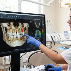
Once the optimal VDO is confirmed, the digital smile design (DSD) process begins. DSD software allows the clinician to overlay the 3D intraoral scans with digital photographs and facial scans of the patient. This creates a virtual patient, enabling the design of the new teeth in perfect harmony with the patient’s facial features, lip line, and phonetic requirements. The proposed design can be 3D printed as a physical mock-up, allowing the patient to “test drive” their new smile directly in their mouth. This functional and aesthetic validation is a critical step before the final CAD/CAM full arch restoration is milled, ensuring that the patient’s expectations are met and that the biomechanical forces are properly distributed[5].
CAD/CAM Dental Lab Technology: Milling Zirconia Monoliths
Computer-aided manufacturing translates digital designs into physical prostheses by precisely milling solid blocks of biocompatible zirconia, ensuring exceptional structural integrity.
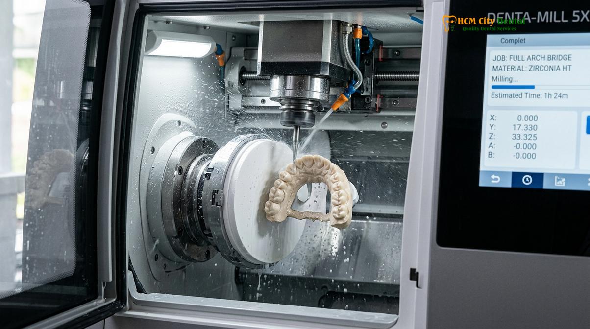
Once the digital design is finalized and approved, the data is sent to a computer-aided manufacturing (CAM) milling machine. This is where the virtual concept becomes a physical reality. For full arch implant-supported restorations, the material of choice is increasingly monolithic zirconia. Unlike traditional porcelain-fused-to-metal (PFM) crowns, which consist of a metal substructure layered with fragile feldspathic porcelain, monolithic zirconia is milled from a single, solid puck of zirconium dioxide.
The zirconia monolithic milling process utilizes advanced 5-axis CNC (Computer Numerical Control) machines. These machines use diamond-coated burs to carve the intricate details of the teeth, gums, and implant connection interfaces with astonishing CAD/CAM precision. Because the restoration is carved from a single block, it possesses immense flexural strength—often exceeding 1,200 Megapascals (MPa). This makes it highly resistant to the heavy occlusal forces generated during chewing and bruxism (teeth grinding)[2].
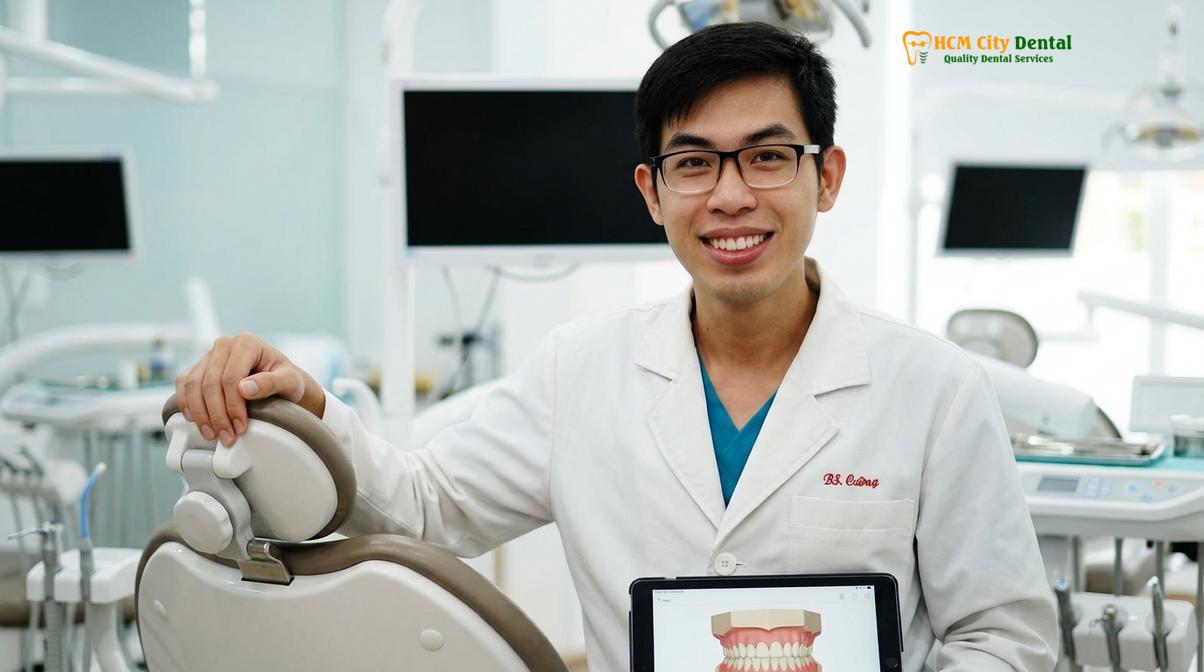
Dr. Nguyen Van Cuong, a leading specialist at HCMC Dental Clinic, emphasizes that the transition to digital dentistry is not just about convenience, but absolute clinical predictability. By integrating intraoral scanning with advanced milling, Dr. Cuong ensures that every Full Mouth Rehabilitation meets the highest international standards for marginal integrity and occlusal harmony. The choice of zirconia is not solely about strength; it is also about biocompatibility. Zirconia is highly bio-inert, meaning it does not provoke an immune response or cause the gingival inflammation sometimes seen with base metal alloys.
Transparent Pricing and Financial Considerations
Understanding the financial investment required for a CAD/CAM full arch restoration is crucial for patients. Clinics catering to international patients often provide structured pricing and significant discounts for advanced booking. Below is an overview of the pricing structure, highlighting the advantages of pre-arrival coordination.
| Implant / Restoration Tier | Standard Walk-in Price (Estimated) | WhatsApp Pre-Booking (-40%) |
|---|---|---|
| Economy (Dentis/Neo Biotech) – Fixture Only | ~$770 (20M VND) | From $460 (12M VND) |
| Standard (Dentium Superline / IBS) – Fixture Only | ~$1,090 (28.3M VND) | From $655 (17M VND) |
| Premium Plus (Straumann BLT-SLA) – Fixture Only | ~$1,795 (46.6M VND) | From $1,080 (28.0M VND) |
| Crown on Implant (Per Tooth) | ~$260 – $385 | From $155 – $230 |
| Full Arch Zirconia Bridge (All-on-4/6) | ~$12,000 – $22,000 per arch | From $7,200 – $13,200 per arch |
| Snap-On Overdenture (Implant-supported) | ~$5,000 – $8,000 per arch | From $3,000 – $4,800 per arch |
In addition to these highly competitive rates, reputable centers offer global manufacturer warranty cards, providing long-term guarantees on implant fixtures. Financial accessibility is further supported through 0% interest monthly installment plans and assistance with international tax offset documentation. Remote post-operative checkups via WhatsApp ensure continuous care for dental tourism patients returning home.
Micro-gap Prevention: Achieving Margin Fit Under 50 Micrometers
A precise margin fit under 50 micrometers is essential to prevent bacterial microleakage and preserve the health of the peri-implant soft tissues.
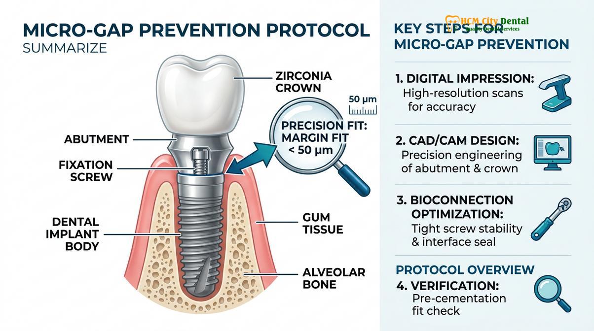
One of the most critical factors determining the long-term success of any dental restoration is the marginal adaptation—the exact point where the restorative material meets the natural tooth structure or the titanium implant abutment. In the analog era, achieving a consistently tight margin was challenging due to the expansion and contraction of impression materials and casting metals. A poor margin fit creates a micro-gap, a microscopic crevice that serves as a harbor for anaerobic bacteria.
Bacterial colonization at the restorative margin leads to microleakage, which can cause recurrent decay under natural tooth crowns or peri-implantitis (bone loss) around dental implants. Furthermore, a bulky or ill-fitting margin violates the biological width—the natural dimension of healthy gingival tissue required to protect the underlying alveolar bone. When the biological width is invaded, the body responds with chronic inflammation and subsequent bone resorption[3].
Clinical Warning: Restorations with a marginal discrepancy greater than 100 micrometers significantly increase the risk of plaque accumulation, gingival inflammation, and eventual failure of the underlying implant or tooth structure.
CAD/CAM technology excels in margin fit precision. High-resolution intraoral scanners capture the exact geometry of the preparation margin, and the 5-axis milling machines can reproduce this geometry with tolerances under 50 micrometers. This level of precision ensures a virtually seamless transition between the zirconia prosthesis and the abutment. This tight seal is paramount for micro-gap prevention, ensuring that the peri-implant tissues remain healthy, pink, and firmly attached to the restoration.
Material Monolithic Strength vs. Traditional Layered Ceramic Chipping
Monolithic zirconia offers superior flexural strength compared to traditional layered ceramics, effectively minimizing the risk of porcelain chipping under heavy bite forces.
For decades, the standard for full arch restorations was the porcelain-fused-to-metal (PFM) bridge. While PFM restorations provided adequate aesthetics, they suffered from a significant biomechanical flaw: the bond between the rigid metal framework and the brittle overlying feldspathic porcelain was prone to failure. Under the immense forces of mastication—especially in patients with parafunctional habits like bruxism—the layered porcelain would frequently fracture or delaminate, a complication known as porcelain chipping.
Repairing a chipped PFM full arch bridge is notoriously difficult and often requires the complete removal and remanufacture of the prosthesis. The introduction of monolithic zirconia has largely addressed this problem. Because the entire restoration is milled from a single, homogenous block of material, there is no weak interface between a substructure and a veneering layer. This porcelain chipping prevention is a massive advantage for implant-supported restorations, which lack the shock-absorbing periodontal ligament found around natural teeth.
“The shift toward monolithic zirconia in full arch implantology has drastically reduced the incidence of catastrophic prosthetic failures, providing patients with a restoration that is highly resilient.”
In the context of All-on-4 or All-on-6 structural biomechanics, the rigidity of monolithic zirconia is highly beneficial. The solid framework distributes occlusal loads evenly across all supporting implants, minimizing the stress concentrated on any single fixture. While some clinicians may choose to add a very thin layer of pink porcelain to simulate gingival tissues (micro-layering), the functional occlusal surfaces remain solid zirconia, ensuring maximum durability and longevity for the CAD/CAM full arch restoration.
Advanced Clinical Protocols: Sedation, Healing, and Full Mouth Rehabilitation
Comprehensive full arch restorations integrate IV conscious sedation for patient comfort and autologous blood concentrates to support tissue healing.
The surgical phase of a full mouth rehabilitation is an extensive procedure that often involves the extraction of failing teeth, alveoloplasty (bone smoothing), guided bone regeneration, and the precise placement of multiple dental implants. To ensure a safe, anxiety-free, and comfortable experience, advanced clinics utilize IV Conscious Sedation. Administered under the supervision of a board-certified anesthesiologist, protocols utilizing specialized medications allow the patient to rest comfortably through the procedure while maintaining their own airway and reflexes. Continuous EKG, blood pressure, and oxygen saturation monitoring ensure absolute safety throughout the surgery.
To optimize the biological response and support healing, clinicians frequently employ Platelet-Rich Fibrin (PRF) therapy. Prior to surgery, a small sample of the patient’s blood is drawn and centrifuged to isolate a concentrated matrix of platelets, white blood cells, and autologous growth factors. This PRF membrane is then placed directly into extraction sockets or mixed with bone grafting materials. The slow release of growth factors stimulates angiogenesis (new blood vessel formation) and significantly supports both soft tissue healing and the osseointegration of the titanium implants[4].
Clinical Case Example: A 62-year-old expatriate presented to HCMC Dental Clinic in Ho Chi Minh City with terminal dentition and severe bite collapse. Utilizing a digital workflow, Dr. Nguyen Van Cuong planned an All-on-6 maxillary reconstruction. Under IV sedation, failing teeth were extracted, PRF was applied, and implants were placed. The patient received a CAD/CAM milled provisional bridge within 48 hours, restoring both function and aesthetics seamlessly.
When to Consult a Specialist for Full Arch Restoration
Recognizing the early signs of bite collapse and terminal dentition is crucial for timely intervention and preventing further structural damage to the jaw.
Patients should consider consulting a prosthodontist or implant specialist if they experience chronic pain in the temporomandibular joint, notice a significant reduction in the lower third of their facial profile, or suffer from advanced periodontal disease that has led to loose or shifting teeth. Delaying treatment for severe tooth loss can result in progressive alveolar bone resorption, making future implant placement more complex and requiring extensive bone grafting procedures. Early consultation allows for a comprehensive 3D digital assessment and the formulation of a predictable, minimally invasive treatment plan.
References
- The Journal of Prosthetic Dentistry. Vertical dimension of occlusion restoration parameters. (2021).
- International Journal of Prosthodontics. Biomechanical load distribution in All-on-4 restorations. (2020).
- Journal of Clinical Periodontology. Biocompatible dental materials and soft tissue response. (2019).
- Clinical Oral Implants Research. Platelet-rich fibrin in guided bone regeneration. (2022).
- Journal of Esthetic and Restorative Dentistry. Digital smile design functional protocols. (2018).
- Vietnam Odonto-Stomatology Association (VOSA). National guidelines on implant-supported prosthodontics and digital dentistry standards. (2023).
Are you experiencing severe tooth loss or bite collapse? Discover how digital dentistry can transform your smile with precision and comfort. Contact HCMC Dental Clinic in Ho Chi Minh City today to schedule your comprehensive 3D scan and consultation for a Full Mouth Rehabilitation.
For customized treatment planning, transparent cost estimates, and direct consultation with Dr. Cuong, visit our comprehensive Full Mouth Rehabilitation service page or message our team directly via WhatsApp to receive an instant assessment.