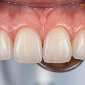
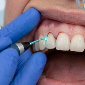
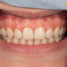
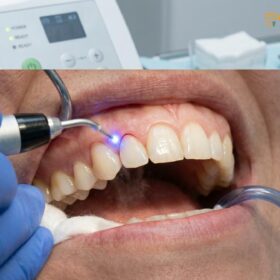
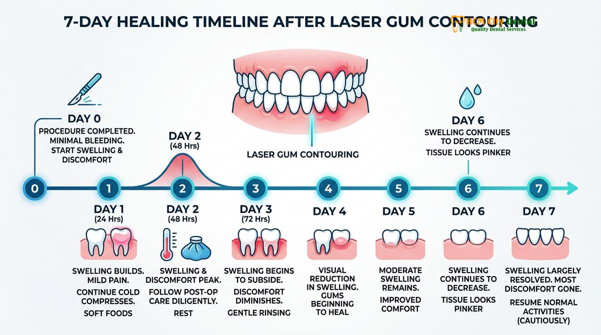
Gum contouring swelling is a natural inflammatory response to surgical tissue modification, typically peaking within 48 hours before gradually subsiding over one to two weeks. Utilizing advanced diode lasers significantly minimizes this edema compared to traditional scalpel methods, ensuring a faster, more comfortable clinical recovery.
Clinical Summary:
Post-operative swelling after gingivectomy or crown lengthening is an expected physiological reaction driven by localized vasodilation and cellular repair mechanisms. The severity and duration of edema depend heavily on the surgical modality. Modern protocols utilizing 810nm diode lasers offer instant coagulation and minimal trauma, drastically reducing recovery time compared to conventional scalpels. Proper adherence to cold compress application, head elevation, and prescribed anti-inflammatory regimens ensures optimal healing, protects the supracrestal tissue attachment, and yields predictable aesthetic outcomes.
Key Takeaways:
- Swelling typically peaks between 48 and 72 hours post-surgery before gradually resolving over the following week.
- AMD Picasso Lasers (810nm) minimize tissue trauma, resulting in significantly less post-operative edema than traditional scalpels.
- Applying cold compresses during the first 24 hours induces vasoconstriction, effectively limiting fluid accumulation.
- Sleeping with the head elevated reduces localized blood pressure in the maxillofacial region, preventing morning swelling.
- Respecting the biological width (minimum 2mm supracrestal tissue attachment) is crucial to prevent chronic inflammation and bone resorption.
Why Do Gums Swell After Surgical Contouring? (Inflammatory Phase)
Swelling is a necessary biological mechanism where increased blood flow delivers leukocytes and healing factors to the surgical site, initiating tissue repair and cellular regeneration.
To understand the clinical timeline of recovery, it is essential to recognize that post-operative edema is not a complication, but rather a highly orchestrated physiological response. When a dental surgeon modifies the gingival architecture—whether to correct a gummy smile or to expose more of the clinical crown—the body immediately registers the micro-trauma. This triggers the release of vasoactive amines, such as histamine and bradykinin, which cause the local capillaries to dilate and become highly permeable.[1]
This increased capillary permeability allows blood plasma, rich in white blood cells (leukocytes), antibodies, and essential nutrients, to extravasate into the interstitial spaces of the gingival tissue. The accumulation of this fluid is what patients visually perceive as swelling. During this acute inflammatory phase, the body is actively working to clear any cellular debris and establish a fibrin network, which serves as the foundational scaffolding for new tissue regeneration.
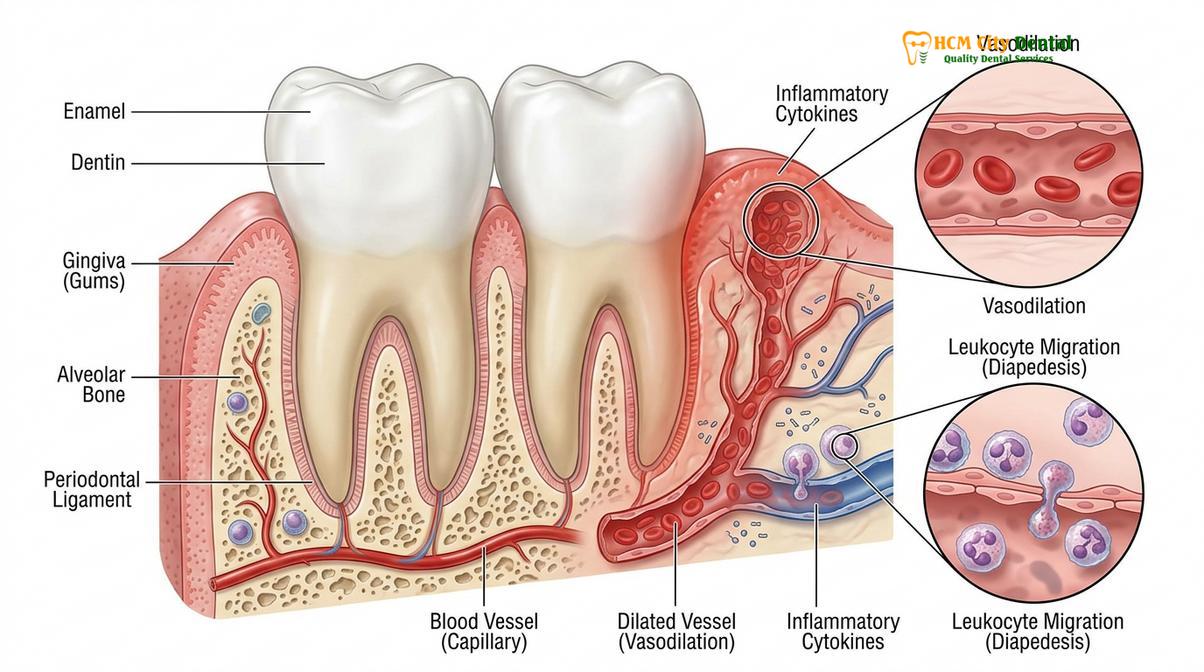
A critical anatomical consideration during this process is the preservation of the biological width, modernly referred to as the supracrestal tissue attachment. This zone, comprising the junctional epithelium and supracrestal connective tissue, requires a minimum of 2mm of space above the alveolar bone crest. If surgical contouring inadvertently invades this space without concurrent osteoplasty (bone reshaping), the body will remain in a state of chronic inflammation, leading to persistent swelling, unpredictable tissue rebound, and potential alveolar bone resorption.
Furthermore, when the dentist calibrates the gingival zenith points—the highest points of the gum scallop, which should be displaced slightly distal to the long axis of the tooth—the precise nature of the incision dictates the extent of the inflammatory response. A clean, minimally invasive approach yields a controlled, transient swelling, whereas excessive tissue manipulation can lead to pronounced post op facial swelling that extends beyond the immediate oral cavity.
Laser vs. Scalpel: How Technology Impacts Post-Operative Edema
Utilizing an 810nm diode laser provides a bloodless, sutureless procedure with instant coagulation, drastically reducing tissue trauma and subsequent swelling compared to traditional scalpels.
The surgical modality chosen for aesthetic gingival reshaping plays the most significant role in determining the severity of post-operative swelling. Historically, gingivectomies were performed using traditional steel scalpels. While effective, scalpel incisions sever blood vessels and nerve endings, leading to intraoperative bleeding, the necessity for sutures, and a robust inflammatory response as the body works to repair the macroscopic tissue damage.
In contemporary aesthetic dentistry, the integration of soft tissue lasers has revolutionized the patient experience. Specifically, the use of AMD Picasso Lasers (Diode soft tissue laser, 810nm) offers unparalleled clinical advantages. The 810nm wavelength is highly absorbed by melanin and hemoglobin, making it exceptionally efficient for soft tissue ablation. As the laser energy vaporizes the excess gingival tissue, it simultaneously cauterizes the adjacent capillaries and seals lymphatic vessels.[2]
“The application of diode lasers in periodontal plastic surgery not only provides a sterile surgical field but also induces photobiomodulation, which downregulates pain receptors and significantly attenuates the acute inflammatory cascade, leading to a rapid 48-hour recovery.”
This instant coagulation means the procedure is virtually bloodless and sutureless. Because the lymphatic vessels are sealed, the extravasation of fluid into the interstitial spaces is profoundly minimized, resulting in a fraction of the swelling typically seen with scalpel surgery. Furthermore, the laser beam is inherently sterile, which drastically minimizes the risk of opportunistic post-operative infections that could otherwise exacerbate inflammation.
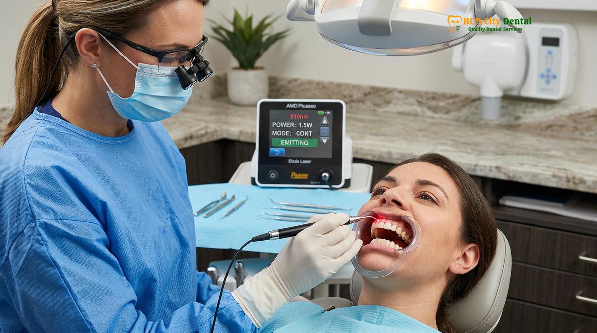
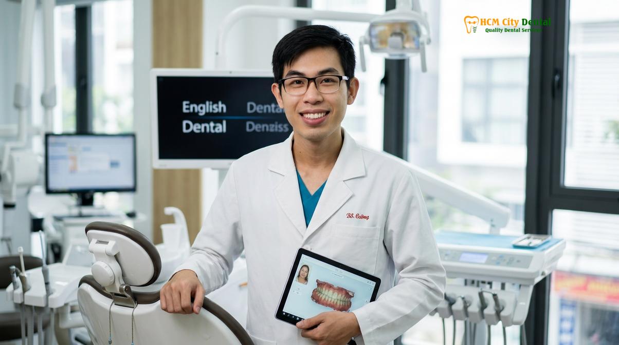
Dr. Nguyen Van Cuong, a leading expert in digital smile design (DSD) and periodontal aesthetics, emphasizes the clinical superiority of laser protocols. From a clinical perspective on zenith calibration, biological width sounding, and laser settings, Dr. Cuong notes that the precision of the AMD Picasso Laser allows for micro-adjustments of the gingival margin without inducing collateral thermal damage to the underlying periosteum or alveolar bone. This meticulous control is the primary reason patients experience minimal discomfort and can often return to their normal routines within days.
Cold Compress Protocols for the First 24 Hours
Applying ice packs to the external cheek in 20-minute intervals induces localized vasoconstriction, effectively limiting fluid extravasation and minimizing acute swelling.
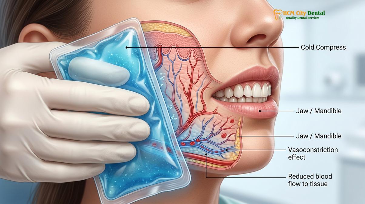
Immediate post-operative care is critical in managing the trajectory of the inflammatory response. The most effective, non-pharmacological intervention during the first 24 hours is the disciplined application of cold therapy. When a cold stimulus is applied to the maxillofacial region, it induces rapid vasoconstriction—the narrowing of blood vessels. This physiological reaction decreases local blood flow and reduces the hydrostatic pressure within the capillaries, thereby limiting the amount of plasma that leaks into the gingival tissues.
To achieve optimal results, patients must follow a strict protocol. The cold compress should be applied to the external cheek or lip area corresponding to the surgical site for 20 minutes, followed by a 20-minute resting period. This intermittent application prevents thermal injury to the skin and avoids a phenomenon known as rebound vasodilation, where the body overcompensates for prolonged cold exposure by forcefully dilating the vessels, which would paradoxically increase swelling.
Clinical Warning: Never apply ice directly to the skin or inside the oral cavity against the healing gums. Always wrap the ice pack in a clean, soft cloth to prevent frostbite and protect the delicate facial epidermis. Discontinue cold therapy after the first 48 hours, as the acute inflammatory phase will have passed.
For patients who have undergone more extensive procedures, such as functional crown lengthening involving bone contouring, utilizing a cold compress jaw surgery protocol is absolutely vital. The cooling effect also slows down cellular metabolism in the affected area, reducing the tissue’s demand for oxygen and minimizing the release of inflammatory mediators. This not only controls the physical distension of the tissues but also provides significant analgesic relief by numbing the local nerve endings.
Elevated Head Sleeping to Minimize Blood Pressure in the Jaws
Keeping the head elevated above the heart during sleep utilizes gravity to prevent fluid pooling in the maxillofacial region, significantly reducing morning edema.
A frequently overlooked aspect of post-operative recovery is the influence of hemodynamics and gravity on tissue swelling. During the day, when a patient is upright, gravity naturally assists in venous return and lymphatic drainage from the head and neck region back toward the heart. However, when a patient lies flat to sleep, this gravitational advantage is lost. The hydrostatic pressure in the maxillofacial capillaries equalizes with the rest of the body, allowing fluids to pool in the surgical sites.
This pooling is the primary reason why patients often notice that their swelling is most pronounced immediately upon waking in the morning. To counteract this, it is clinically advised to practice head elevated sleep gums protocols for the first three to five nights following the procedure. By propping the head up with two or three pillows, or by sleeping in a recliner at a 30 to 45-degree angle, the patient maintains the gravitational gradient necessary to facilitate continuous fluid drainage.
This simple postural adjustment significantly reduces the localized blood pressure in the jaws, preventing the exacerbation of edema. It also minimizes the throbbing sensation that some patients experience when lying flat, as the reduced vascular pressure translates to less mechanical stress on the sensitized nerve endings surrounding the surgical site.
Anti-inflammatory Medications and Vitamin Supplements to Support Healing
A targeted pharmacological approach combining NSAIDs with cellular-supporting vitamins accelerates the resolution of edema and promotes robust soft tissue regeneration.
While physical modalities like cold compresses and head elevation are crucial, pharmacological management plays an equally important role in modulating the inflammatory cascade. Non-Steroidal Anti-Inflammatory Drugs (NSAIDs), such as Ibuprofen, are typically the first line of defense. These medications work by inhibiting the cyclooxygenase (COX) enzymes, thereby blocking the synthesis of prostaglandins—lipid compounds that are primary drivers of inflammation, pain, and swelling.[3]
For optimal efficacy, NSAIDs should be taken on a strict schedule as prescribed by the dental surgeon, rather than on an “as-needed” basis, to maintain a consistent therapeutic level in the bloodstream during the peak inflammatory window (the first 48 to 72 hours). This proactive approach is highly effective to reduce gum inflammation before it reaches its maximum potential.
Beyond analgesics, nutritional support is vital for cellular repair. Vitamin C (Ascorbic Acid) is a critical cofactor in the synthesis of collagen, the primary structural protein in gingival tissue. Adequate Vitamin C levels ensure that the new collagen fibrils cross-link correctly, providing strength and elasticity to the healing gums. Additionally, Omega-3 fatty acids, found in fish oil supplements, possess natural anti-inflammatory properties that can synergistically support the reduction of post-operative edema.
Advanced Clinical Workflows and Surgical Partnerships
For complex skeletal discrepancies causing a gummy smile, multidisciplinary orthognathic surgery combined with precise soft tissue contouring ensures optimal functional and aesthetic harmony.
While laser gingivectomy is highly effective for patients with excess gingival display due to altered passive eruption or gingival hyperplasia, it is not a universal solution for all gummy smiles. Comprehensive diagnostics, including cephalometric analysis and digital smile design, are required to determine the true etiology of the aesthetic concern. In some instances, the excessive gingival display is not a soft tissue issue, but rather a skeletal discrepancy known as Vertical Maxillary Excess (VME).
In cases of severe VME, simply removing gum tissue would violate the biological width, expose the root surfaces, and fail to address the underlying skeletal disproportion. For these complex cases, a multidisciplinary approach is required. This involves orthognathic surgery, specifically a Le Fort I osteotomy, where the entire upper jaw is surgically repositioned superiorly to reduce the vertical height of the maxilla.[4]
Clinical Case Review: A 28-year-old patient presented to HCMC Dental Clinic in Ho Chi Minh City with a severe gummy smile (8mm of gingival display). Clinical diagnostics revealed a combination of a hyperactive levator muscle and mild Vertical Maxillary Excess. Instead of invasive jaw surgery, the multidisciplinary team, guided by Dr. Nguyen Van Cuong, executed a combined approach: a laser gingivectomy to establish ideal zenith points, followed by Surgical Lip Repositioning to restrict the upward pull of the upper lip. The result was a harmonious, balanced smile achieved with minimally invasive protocols.
For severe skeletal cases requiring Le Fort I osteotomy orthognathic surgery, the clinic works in collaboration with maxillofacial specialists at the National Hospital of Odonto-Stomatology in Ho Chi Minh City (Bệnh viện Răng Hàm Mặt Trung Ương TP.HCM), the highest clinical authority in the region. This surgical jaw partnership ensures that patients receive the safest, most advanced care possible, bridging the gap between aesthetic dentistry and major maxillofacial reconstruction.
Transparent Pricing and Dental Tourism in Ho Chi Minh City
HCMC Dental Clinic offers world-class, laser-assisted periodontal treatments at highly competitive rates, making it a premier destination for international patients seeking aesthetic excellence.
The globalization of healthcare has positioned Vietnam as a leading hub for high-quality, cost-effective medical and dental care. Dental tourism in Ho Chi Minh City is thriving, driven by the availability of state-of-the-art technology, internationally trained specialists, and transparent, highly competitive pricing structures. Patients traveling from Australia, the United States, and Europe can access premium aesthetic procedures at a fraction of the cost in their home countries, without compromising on clinical standards or safety protocols.
Prior to any surgical intervention, a comprehensive foundation of oral health must be established. This typically begins with professional dental scaling and root planing to eliminate calculus and bacterial biofilm, ensuring the surgical site is free from active periodontal disease. Once the tissues are healthy, the aesthetic phase can commence.
According to the latest clinic fee schedule, the pricing structure for comprehensive aesthetic gum contouring procedures is designed to be transparent and accessible. Patients who utilize the digital booking system can take advantage of a significant -40% WhatsApp booking discount.
| Clinical Procedure | Standard Walk-in Fee (VND) | Discounted Fee (-40% WhatsApp) | Estimated USD |
|---|---|---|---|
| Laser Gingivectomy (per tooth) | 2,500,000 VND | 1,500,000 VND | ~$60 USD |
| Full-Arch Laser Gum Contouring (up to 10 teeth) | 20,000,000 VND | 12,000,000 VND | ~$480 USD |
| Functional/Esthetic Crown Lengthening (per tooth, incl. bone contouring) | 5,000,000 VND | 3,000,000 VND | ~$120 USD |
| Surgical Lip Repositioning / Lip Lowering | 25,000,000 VND | 15,000,000 VND | ~$600 USD |
| Botox Gummy Smile Correction (both sides) | 7,500,000 VND | 4,500,000 VND | ~$180 USD |
Whether a patient requires a simple laser adjustment of the gingival margins or a complex functional crown lengthening involving osteotomy and osteoplasty, the clinical team provides a detailed, itemized treatment plan during the initial consultation. This commitment to transparency ensures that patients can make informed decisions regarding their aesthetic investments.
When to See a Doctor
While mild to moderate swelling, tenderness, and minor oozing are expected physiological responses during the first 48 to 72 hours, certain clinical signs warrant immediate professional evaluation. It is crucial to differentiate between normal inflammatory healing and potential post-operative complications.[5]
You should contact your dental surgeon immediately if you experience any of the following red flags:
- Progressive Swelling: Edema that continues to worsen after the fourth day, rather than subsiding, or swelling that rapidly extends to the neck or under the jaw, potentially compromising the airway.
- Signs of Infection: The presence of purulent discharge (pus) from the surgical site, a foul odor or taste in the mouth, or localized tissue that turns dark red, purple, or black (necrosis).
- Systemic Symptoms: A persistent fever above 101°F (38.3°C), chills, or swollen, tender lymph nodes in the neck.
- Uncontrolled Bleeding: Active, bright red bleeding that does not stop after applying firm, continuous pressure with a sterile gauze pad for 20 to 30 minutes.
- Severe, Intractable Pain: Pain that is sharp, throbbing, and unresponsive to the prescribed maximum doses of analgesic medications.
Early intervention is key to managing complications. If you have any doubts about your recovery trajectory, it is always safer to seek a clinical assessment to ensure your healing remains on track.
Frequently Asked Questions
When does swelling peak after gum contouring?
Swelling typically peaks between 48 and 72 hours following the surgical procedure before gradually subsiding. Adhering to cold compress protocols and keeping your head elevated during this critical window significantly minimizes the maximum extent of the edema.
Can warm compresses help?
Warm compresses should only be applied after the first 72 hours have passed and the acute inflammatory phase has stabilized. Applying heat too early induces vasodilation, which increases blood flow and exacerbates swelling and post-operative discomfort.
Does ice help gums recover faster?
Yes, applying ice packs externally during the first 24 hours induces localized vasoconstriction, which limits fluid accumulation and reduces cellular metabolic demand. This effectively minimizes tissue distension, allowing the gingival architecture to heal more efficiently.
How long should I sleep with my head elevated?
It is clinically recommended to sleep with your head elevated above your heart for the first three to five nights post-surgery. This utilizes gravity to promote venous return and lymphatic drainage, preventing morning facial edema.
Is it normal for the gums to look white during recovery?
Yes, a white or yellowish film over the surgical site is a normal physiological response representing the formation of a fibrin clot. This acts as a natural biological bandage protecting the underlying healing tissue and should not be scrubbed off.
References
- Journal of Clinical Periodontology. Inflammatory responses following periodontal plastic surgery. (2021).
- International Journal of Periodontics & Restorative Dentistry. Diode laser vs scalpel gingivectomy: Healing outcomes. (2020).
- Journal of Aesthetic and Restorative Dentistry. Biological width and supracrestal tissue attachment around restorations. (2019).
- Journal of Oral and Maxillofacial Surgery. Le Fort I osteotomy stability in vertical maxillary excess. (2022).
- Clinical Oral Investigations. Post-operative edema management in oral surgery. (2018).