Gum contouring recovery typically spans 48 hours to two weeks, depending on whether a modern diode laser or a traditional scalpel is utilized. Patients can expect minor swelling and mild sensitivity during the initial days, which resolves rapidly with proper oral hygiene and a temporary soft food diet.
Clinical Summary:
Recovery from gum contouring is a highly predictable biological process, especially when utilizing advanced 810nm diode laser technology. The healing timeline involves immediate cauterization, followed by cellular proliferation and tissue maturation over 7 to 14 days. Maintaining the biological width and adhering to strict post-operative protocols—such as a cold, soft diet and modified brushing techniques—ensures optimal esthetic outcomes without compromising the supracrestal tissue attachment. Clinical oversight is essential to monitor zenith point stabilization and prevent infection.
Key Takeaways:
- Laser-assisted contouring significantly reduces bleeding and accelerates the healing timeline compared to traditional scalpel methods.
- The first 72 hours require a strict cold and soft diet to prevent clot disruption and minimize inflammation.
- Gentle oral hygiene practices must be resumed by day four to prevent plaque accumulation around the healing margins.
- Full tissue maturation and final esthetic stabilization of the gingival zenith points can take up to several weeks.
- Severe skeletal gummy smiles may require collaborative orthognathic surgery for comprehensive correction.
The Soft Tissue Healing Process: What Happens to Gums Cells
The biological healing of gingival tissue involves a complex cascade of hemostasis, cellular proliferation, and remodeling. Utilizing advanced laser technology minimizes trauma, allowing epithelial cells to migrate rapidly while preserving the critical supracrestal tissue attachment.
Understanding the physiological mechanisms behind the healing timeline gum reshaping requires a deep dive into cellular biology and periodontal anatomy. When gingival tissue is modified, the body immediately initiates a highly orchestrated wound-healing response. The primary goal of this response is to re-establish the epithelial barrier and protect the underlying alveolar bone from bacterial invasion. The success of this process relies heavily on the surgical technique employed and the precise management of the biological width.
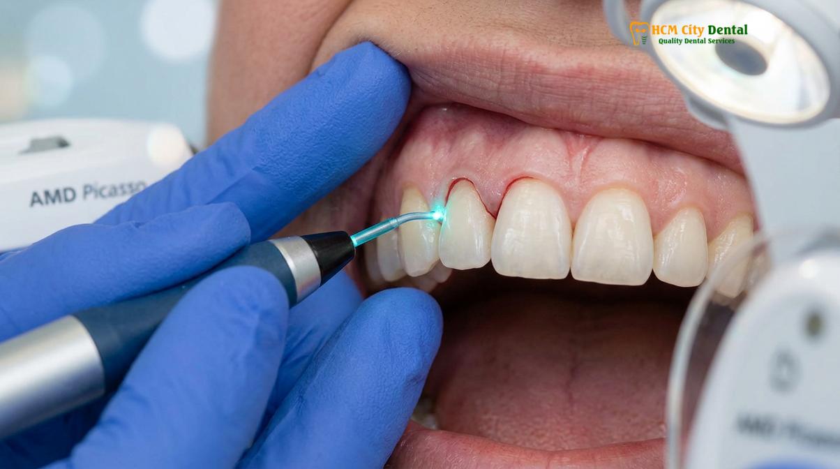
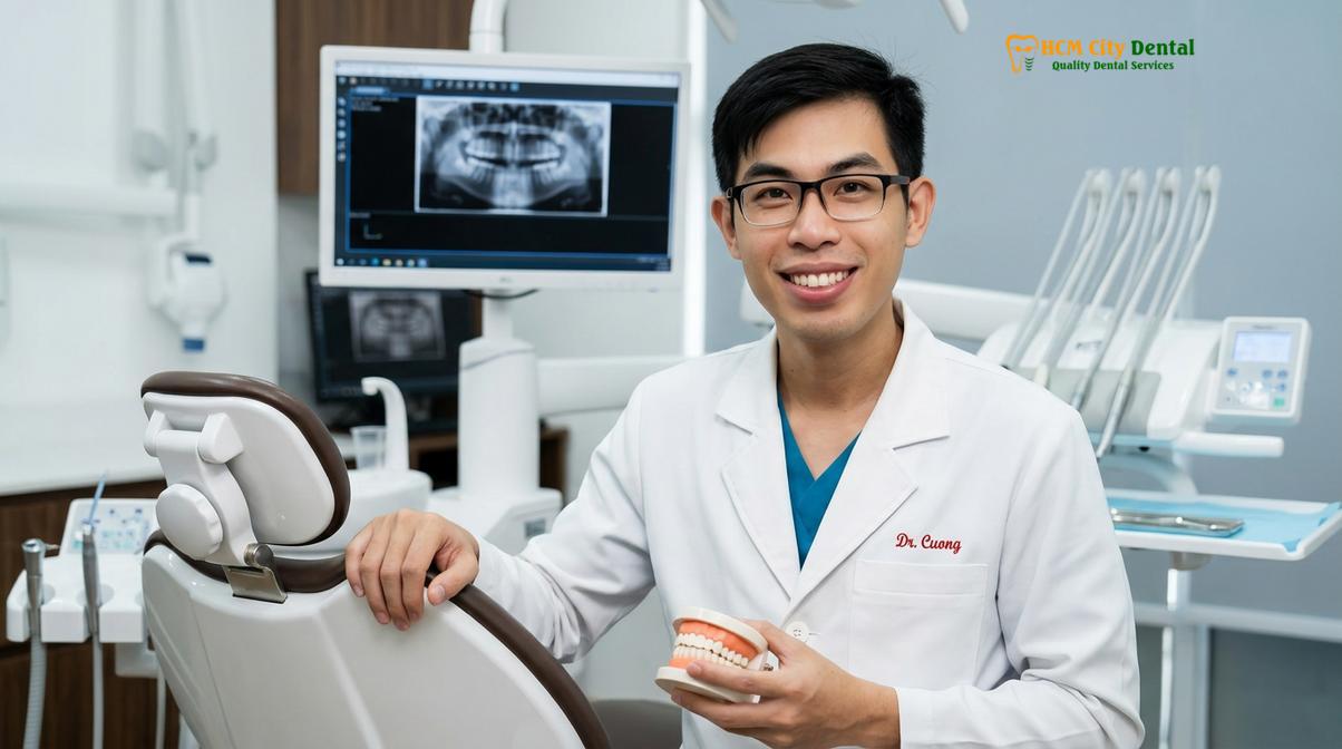
At the forefront of modern periodontal plastic surgery is the utilization of AMD Picasso Lasers. These advanced diode soft tissue lasers operate at an 810nm wavelength, which is highly absorbed by soft tissue chromophores such as melanin and hemoglobin. When Dr. Nguyen Van Cuong performs a gingivectomy, the clinical perspective on zenith calibration and biological width sounding is paramount. The laser energy vaporizes the targeted intracellular fluid, resulting in precise tissue ablation while simultaneously sealing capillaries and lymphatic vessels. This photothermal effect creates a virtually bloodless surgical field, eliminating the need for sutures and drastically reducing post-operative edema.
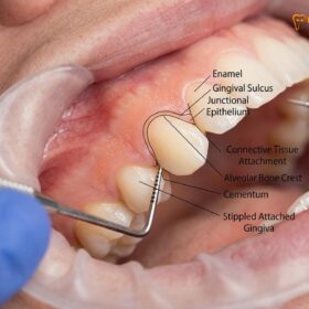
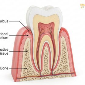
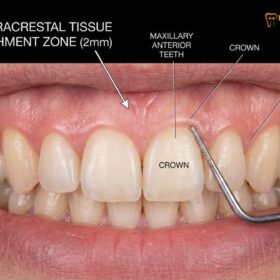
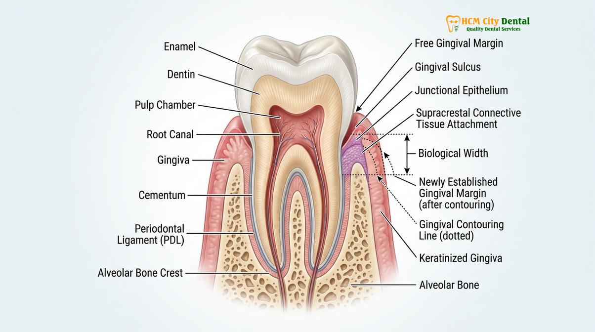
The preservation of the supracrestal tissue attachment (formerly known as the biological width) is a non-negotiable clinical parameter. This attachment consists of approximately 1mm of junctional epithelium and 1mm of connective tissue attachment above the alveolar bone crest. If the contouring process violates this 2mm zone without concurrent osseous recontouring (osteoplasty or osteotomy), the body will respond with chronic inflammation, unpredictable tissue rebound, or pathological bone resorption as it attempts to re-establish the necessary biological space [1].
“The application of 810nm diode lasers in periodontal plastic surgery not only provides a bloodless surgical field but also creates a photobiomodulation effect that significantly reduces post-operative pain and accelerates the initial epithelialization phase.”
Following the immediate hemostasis achieved by the laser, the tissue enters the proliferative phase. Fibroblasts migrate into the surgical site to synthesize new collagen, while epithelial cells begin to bridge the wound margins at a rate of approximately 0.5mm per day. Because the laser beam sterilizes the surgical margin as it cuts, the risk of secondary bacterial infection is profoundly minimized, allowing for a rapid 48-hour initial recovery phase that is vastly superior to the secondary intention healing required after traditional scalpel incisions [2].
Days 1-3: Managing Minor Soreness and the Cold Food Diet
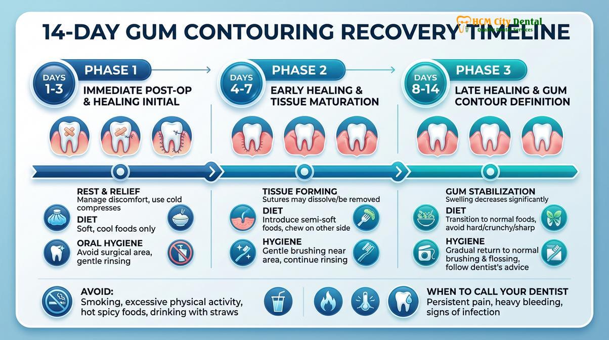
The initial 72 hours post-surgery are critical for stabilizing the surgical site and managing mild inflammatory responses. Patients must strictly adhere to a cold, soft diet and avoid mechanical trauma to ensure the initial fibrin network remains intact.
The immediate post-operative period is characterized by the body’s acute inflammatory response, even when minimally invasive laser protocols are utilized. Patients typically experience mild tenderness, slight tissue erythema (redness), and minor localized swelling. Because the diode laser cauterizes nerve endings during the procedure, acute pain is exceedingly rare; most patients find that over-the-counter analgesics, such as acetaminophen or ibuprofen, are more than sufficient to manage any discomfort.
A critical component of this early recovery phase is the implementation of a strict soft diet after gum surgery. The mechanical forces generated during mastication (chewing) can easily disrupt the fragile fibrin clot that forms over the surgical site. Furthermore, thermal regulation is vital. Consuming hot foods or beverages induces vasodilation—the widening of blood vessels—which can dissolve the clot, restart bleeding, and exacerbate swelling.
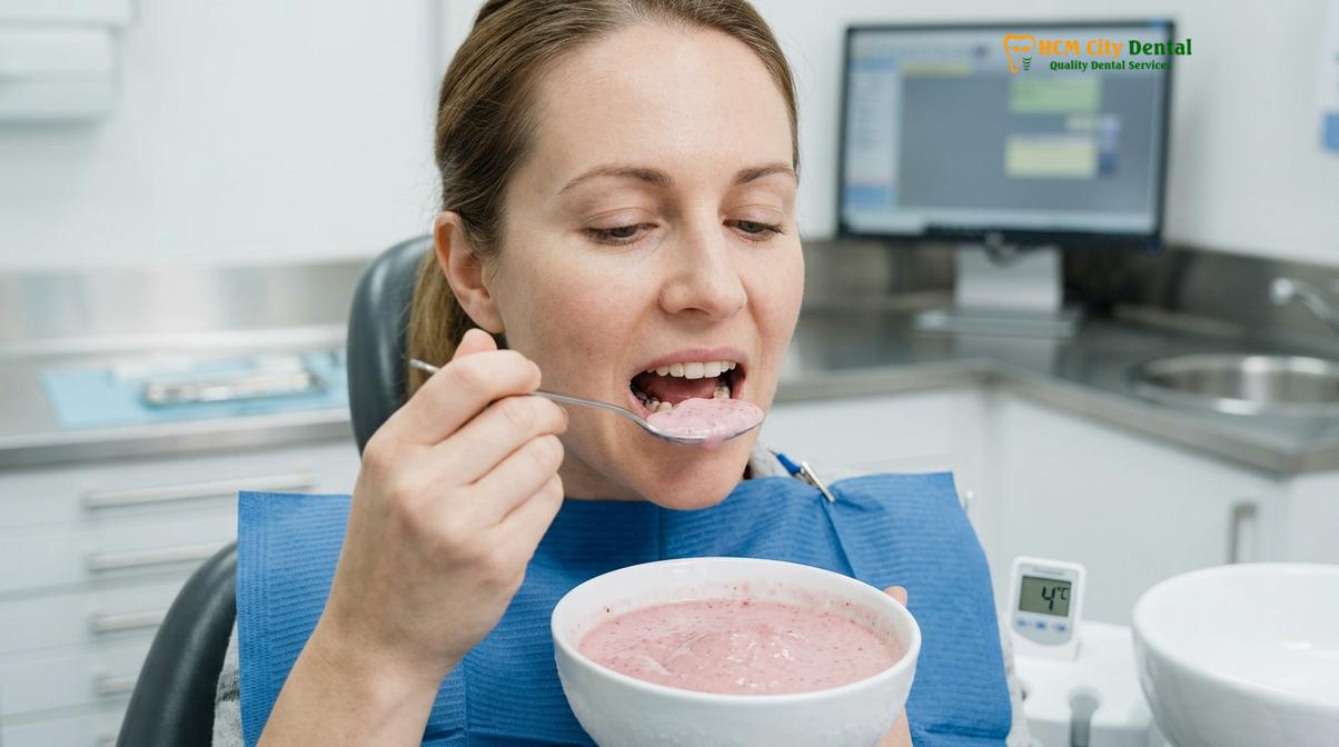
During Days 1 to 3, patients should consume foods that require zero chewing and are served cold or at room temperature. Excellent dietary choices include:
- Chilled protein shakes and nutritional smoothies (consumed without a straw to prevent negative intraoral pressure).
- Cold Greek yogurt, which provides essential proteins necessary for collagen synthesis.
- Applesauce and pureed fruit blends.
- Chilled gazpacho or room-temperature blended soups.
- Soft scrambled eggs allowed to cool completely.
It is imperative to avoid any physical exertion or heavy lifting during this 72-hour window. Elevated cardiovascular activity increases systemic blood pressure, which can lead to throbbing sensations at the surgical site and potential delayed hemorrhage. Resting with the head slightly elevated on extra pillows can further assist in minimizing fluid accumulation and facial edema.
Days 4-7: Resuming Gentle Brushing and Flossing Workflows
As the initial inflammatory phase subsides, reintroducing modified mechanical plaque control is essential to prevent bacterial colonization. Patients should utilize ultra-soft bristles and specialized antimicrobial rinses while avoiding direct friction on the healing margins.
By the fourth day, the acute inflammatory phase transitions into the proliferative phase, where active gums tissue repair is underway. The surgical margins begin to re-epithelialize, forming a delicate new layer of mucosal tissue. While the risk of bleeding significantly decreases, the new tissue remains highly susceptible to mechanical abrasion. Therefore, modifying your oral hygiene after gingivectomy is crucial to prevent plaque-induced inflammation without traumatizing the healing site.
Standard brushing techniques must be temporarily abandoned. Patients are instructed to use an ultra-soft surgical toothbrush. The bristles should only contact the occlusal (biting) surfaces and the coronal portions of the teeth, strictly avoiding the cervical margins where the gums meet the teeth. The modified Bass technique or a gentle roll technique directed away from the gingival margin is recommended to remove biofilm safely.
Because mechanical brushing is limited, chemical plaque control becomes the primary defense against bacterial accumulation. Clinical protocols often involve the prescription of a 0.12% chlorhexidine gluconate mouthrinse. This broad-spectrum antimicrobial agent binds to oral tissues and provides substantivity, meaning it continues to release its antibacterial properties for up to 12 hours after rinsing. Patients should gently bathe the surgical site with the rinse rather than swishing vigorously, allowing the fluid to fall out of the mouth passively into the sink.
Clinical Case Observation: HCMC Dental Clinic
A 28-year-old patient visited HCMC Dental Clinic in Ho Chi Minh City presenting with altered passive eruption, resulting in short clinical crowns and a prominent gummy smile. Dr. Nguyen Van Cuong performed a full-arch laser gingivectomy utilizing digital smile design to calibrate the zenith points. By Day 5 of the recovery protocol, the patient exhibited zero bleeding on probing, complete cessation of mild tenderness, and excellent initial epithelialization, allowing for a seamless transition back to a normal professional speaking schedule.
Flossing should be resumed with extreme caution. The floss must be carefully guided through the interproximal contacts and moved slightly subgingivally without snapping or forcing the thread against the healing papillae. If flossing causes any discomfort or pinpoint bleeding, it should be suspended for an additional 48 hours.
Foods to Avoid to Prevent Irritation and Delayed Healing
Certain dietary choices can mechanically disrupt healing tissues, induce thermal trauma, or trigger chemical irritation at the surgical site. Avoiding spicy, crunchy, acidic, and excessively hot foods is paramount to preventing complications.
The trajectory of your recovery is heavily influenced by dietary compliance. While the soft tissue laser accelerates the biological timeline, the newly formed epithelium remains fragile for up to two weeks. Introducing inappropriate foods can cause mechanical lacerations, chemical burns, or thermal shock, all of which prolong the inflammatory phase and delay final tissue maturation.
Mechanically Traumatic Foods: Foods with sharp edges or hard textures must be strictly avoided. This includes potato chips, crusty bread, popcorn, nuts, and hard candies. A single sharp fragment of a tortilla chip can easily lacerate the healing gingival margin, requiring the body to restart the epithelialization process from the beginning.
Chemically Irritating Foods: Spicy foods containing capsaicin induce a localized neurogenic inflammatory response, causing severe burning sensations and vasodilation at the surgical site. Similarly, highly acidic foods—such as citrus fruits, tomatoes, and vinegar-based dressings—lower the oral pH. This acidic environment not only causes acute stinging but can also denature the delicate proteins forming the new tissue matrix.
Thermally Traumatic Foods: As previously noted, hot soups, boiling teas, and freshly cooked meats must be avoided. Thermal trauma can cause the newly formed capillaries to rupture. Furthermore, patients should avoid using drinking straws; the negative intraoral pressure generated by suction can dislodge forming clots, particularly if the procedure involved deeper osseous recontouring.
Advanced Clinical Workflows: From Lasers to Orthognathic Surgery
While minor gingival discrepancies are efficiently managed with soft tissue lasers, complex skeletal cases demand a multidisciplinary surgical approach. Comprehensive treatment planning integrates digital smile design, osseous recontouring, and occasionally orthognathic surgery.
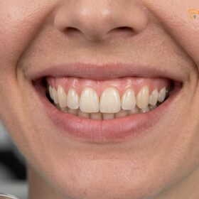
The etiology of a gummy smile (excessive gingival display) is multifactorial, and accurate clinical diagnosis is the cornerstone of successful treatment. While altered passive eruption or minor gingival hyperplasia can be beautifully corrected with the AMD Picasso Diode laser, other anatomical factors require more advanced surgical interventions to achieve lasting dentofacial harmony.
When the underlying issue involves a hyperactive levator labii superioris muscle—the muscle responsible for elevating the upper lip—the lip raises excessively during a full smile, exposing a wide band of gingiva. In these instances, laser contouring alone is insufficient. The clinical workflow may incorporate Botox injections to temporarily paralyze the hyperactive muscle, or a surgical lip repositioning procedure. Lip lowering surgery involves removing a strip of mucosa from the maxillary vestibule and suturing the lip in a lower, more restrictive position, thereby permanently reducing gingival display [5].
“Achieving a harmonious gingival architecture requires meticulous evaluation of the osseous crest; failure to respect the supracrestal tissue attachment inevitably leads to chronic marginal inflammation and unpredictable tissue rebound.”
In cases of severe skeletal discrepancies, specifically Vertical Maxillary Excess (VME), the entire upper jaw bone has grown too far downward. For these complex skeletal cases, HCMC Dental Clinic relies on its Surgical Jaw Partners. The clinic works in close collaboration with elite maxillofacial specialists at the National Hospital of Odonto-Stomatology in Ho Chi Minh City (Bệnh viện Răng Hàm Mặt Trung Ương TP.HCM), the highest clinical authority in the region. These cases require a Le Fort I osteotomy—a major orthognathic surgery where the maxilla is sectioned, impacted upward, and stabilized with titanium plates to fundamentally alter the facial skeleton [4].
Regardless of the surgical modality, Dr. Cuong emphasizes the importance of Digital Smile Design (DSD). Before any tissue is altered, digital calipers and specialized software are used to map the ideal gingival zenith points—the highest point of the gum scallop, which should be displaced slightly distal to the long axis of the tooth. This meticulous pre-operative planning ensures that the final biological contours perfectly complement the patient’s facial aesthetics.
Comprehensive Cost Guide for Gum Contouring Procedures
Treatment costs vary based on the complexity of the procedure, ranging from simple laser gingivectomies to comprehensive functional crown lengthening and skeletal corrections. Transparent pricing ensures patients can make informed decisions.
Investing in periodontal plastic surgery requires an understanding of the specific techniques required for your unique anatomy. The integration of advanced laser technology and digital planning provides superior outcomes, and the pricing structure reflects the clinical expertise and equipment utilized. For international patients engaging in dental tourism, Ho Chi Minh City offers world-class periodontal care at a fraction of Western costs.
According to the latest clinic fee schedule, the pricing parameters for esthetic gum procedures are structured as follows:
| Clinical Procedure | Standard Walk-in Fee (VND) | Online Booking Fee (VND) | Estimated USD |
|---|---|---|---|
| Laser Gingivectomy (per tooth) | 2,500,000 | 1,500,000 | ~$60 |
| Full-Arch Laser Gum Contouring (up to 10 teeth) | 20,000,000 | 12,000,000 | ~$480 |
| Functional/Esthetic Crown Lengthening (per tooth, includes bone contouring) | 5,000,000 | 3,000,000 | ~$120 |
| Surgical Lip Repositioning / Lip Lowering | 25,000,000 | 15,000,000 | ~$600 |
| Botox Gummy Smile Correction (both sides) | 7,500,000 | 4,500,000 | ~$180 |
Patients are highly encouraged to utilize the -40% WhatsApp booking discount to secure the online rates listed above. Comprehensive treatment planning, including 3D imaging and digital smile design, is conducted during the initial consultation to determine whether a simple soft tissue laser approach is sufficient, or if osseous crown lengthening is required to respect the biological width [3]. For more detailed information on candidacy and procedural workflows, explore our comprehensive guide on Gum Contouring & Gummy Smile.
When to See a Doctor: Identifying Post-Operative Complications
While the use of sterile diode lasers makes post-operative complications exceedingly rare, patients must remain vigilant during the healing timeline. Differentiating between normal biological healing and pathological complications is crucial for maintaining the integrity of the surgical results. Normal healing includes mild, localized swelling, slight tissue blanching or a white fibrinous slough over the surgical site (which is often mistaken for pus but is actually normal protein coagulation), and minor sensitivity to temperature changes.
Immediate Clinical Attention is Required If You Experience:
- Uncontrolled Hemorrhage: Bleeding that does not stop after 20 minutes of continuous, firm pressure with a damp gauze pad.
- Purulent Exudate: The presence of thick, yellow or green pus discharging from the gingival margins, indicating an active bacterial infection.
- Systemic Symptoms: A fever exceeding 101°F (38.3°C), chills, or swollen lymph nodes in the neck and jaw.
- Severe, Throbbing Pain: Pain that progressively worsens after the third day and is unresponsive to prescribed analgesic medications.
If any of these warning signs manifest, it is imperative to contact the clinic immediately for a thorough clinical evaluation. Early intervention with targeted antibiotic therapy or localized debridement can swiftly resolve infections and protect the esthetic outcome of the contouring procedure.
Frequently Asked Questions
Can I eat hot food after gum contouring?
No, you should strictly avoid hot foods and beverages for at least the first 48 to 72 hours following your procedure. High temperatures cause vasodilation, which expands blood vessels and can dissolve the initial blood clot, leading to secondary bleeding and increased inflammation. Stick to a cold or room-temperature soft diet during this critical initial healing phase.
When can I return to hard chewing?
Most patients can safely return to a normal diet, including harder foods, after 10 to 14 days, provided the gingival margins have adequately re-epithelialized. However, if your procedure involved osseous recontouring (bone removal), you may need to wait up to three weeks before chewing highly crunchy or tough foods to prevent mechanical trauma to the healing attachment.
How do I clean my teeth during the first week?
During the first week, you must use an ultra-soft surgical toothbrush and avoid direct contact with the healing gum margins. Brush only the biting surfaces of your teeth and use a prescribed antimicrobial mouthwash, such as 0.12% chlorhexidine, or a warm saline rinse to chemically control plaque without mechanically disrupting the delicate new tissue.
Will my gums grow back after the procedure?
When performed correctly with proper respect for the biological width and necessary bone contouring, gum tissue will not grow back. However, if only soft tissue is removed without adjusting the underlying bone in cases where the supracrestal attachment is violated, the tissue is highly likely to rebound to its original position due to biological memory.
Is the laser recovery faster than traditional scalpel surgery?
Yes, recovery following diode laser contouring is significantly faster and more comfortable than traditional scalpel surgery. The 810nm laser instantly cauterizes blood vessels and nerve endings, resulting in a bloodless field, zero need for sutures, minimal post-operative swelling, and a rapid 48-hour initial healing phase.
References
- Journal of Clinical Periodontology. Esthetic crown lengthening outcomes and biological width. (2021).
- International Journal of Periodontics & Restorative Dentistry. Diode laser vs scalpel gingivectomy healing. (2020).
- Journal of Aesthetic and Restorative Dentistry. Supracrestal tissue attachment and restorative margins. (2019).
- Journal of Oral and Maxillofacial Surgery. Le Fort I osteotomy stability in vertical maxillary excess. (2022).
- Clinical Oral Investigations. Surgical lip repositioning longevity and patient satisfaction. (2018).