Crown lengthening for veneers in HCMC is a precise periodontal procedure that reshapes gingival tissue and underlying bone to expose more healthy tooth structure. This clinical step ensures optimal aesthetic proportions and provides a stable, healthy foundation for durable porcelain restorations without violating the biological width.
Clinical Summary:
Crown lengthening is a critical preparatory phase for both cosmetic and restorative dentistry. By utilizing advanced diode lasers and precise osteotomy techniques, clinicians adjust the gingival zenith points and supracrestal tissue attachment. This meticulous process prevents chronic inflammation, ensures proper veneer seating, and resolves complex structural issues like subgingival decay or fractured teeth. When executed correctly, it establishes a harmonious, long-lasting smile architecture, making it an indispensable step before finalizing high-end porcelain restorations.
Key Takeaways:
- Requires a minimum of 2mm supracrestal tissue attachment to maintain long-term periodontal health.
- Utilizes AMD Picasso Lasers (810nm) for bloodless, sutureless, and highly precise tissue contouring.
- Essential for treating deep subgingival decay and salvaging severely fractured teeth.
- Prevents biological width violations that commonly cause chronic gum inflammation and bone loss.
- Often integrated with Digital Smile Design (DSD) to calibrate perfect gingival zenith points for veneers.
What is Functional Crown Lengthening and Why is it Necessary?
Functional crown lengthening is a surgical periodontal procedure that lowers the gum and bone levels to expose healthy tooth structure, creating a solid, disease-free foundation for restorative dental work.
In the realm of comprehensive restorative dentistry, the structural integrity of the foundation is just as critical as the aesthetic appeal of the final restoration. Functional crown lengthening is distinct from purely aesthetic gum contouring. While aesthetic contouring primarily addresses the soft tissue to correct a gummy smile or uneven gum lines, functional lengthening involves the meticulous reshaping of both the gingival tissue and the underlying alveolar bone. This procedure is necessitated when a tooth is severely compromised by decay, trauma, or wear, leaving insufficient clinical crown height to support a durable restoration such as a porcelain veneer or a full-coverage crown[1].
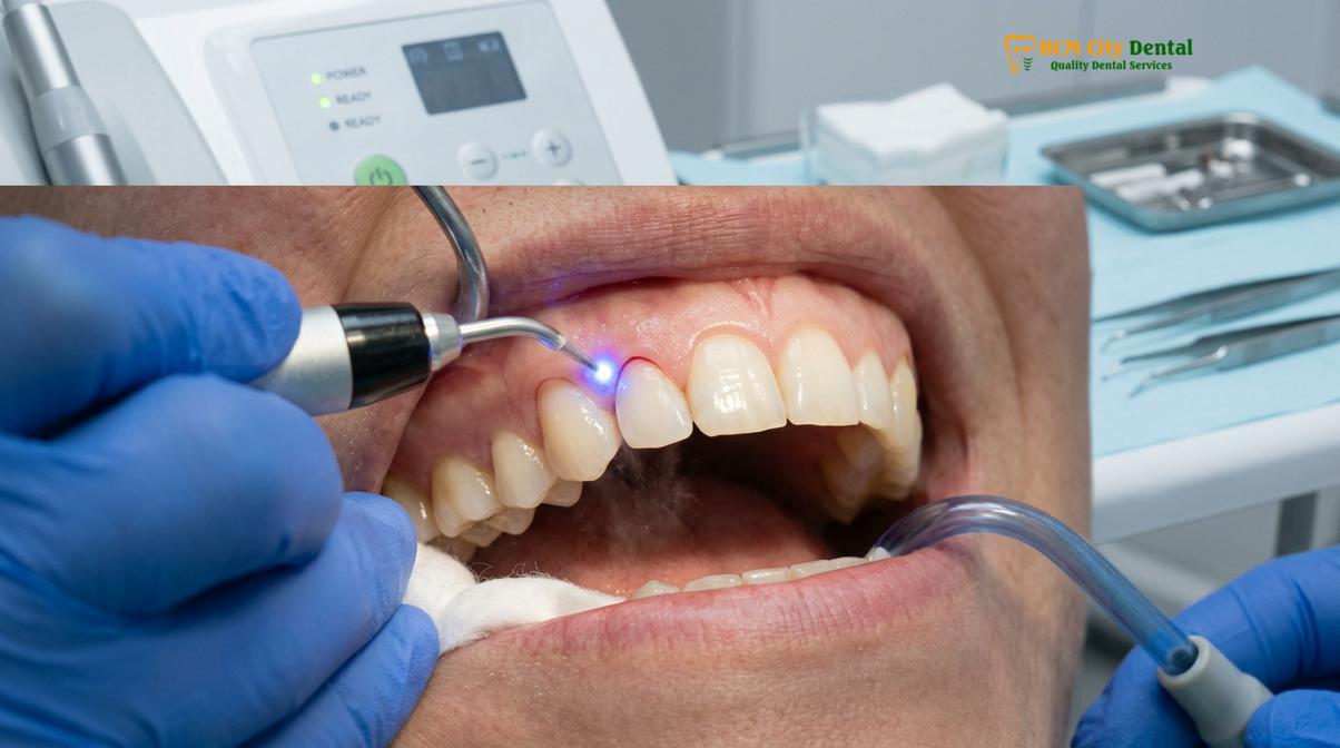
The primary objective of this procedure is to expose enough sound tooth structure to achieve a proper “ferrule effect.” A ferrule is a band of restorative material that completely encircles the external dimension of the residual tooth structure. Clinical guidelines dictate that a minimum of 1.5 to 2.0 millimeters of healthy tooth structure must be exposed above the bone level to ensure the mechanical retention and structural resistance of the final crown or veneer. Without this critical ferrule, the restoration is highly susceptible to decementation, and the underlying tooth root is at a significantly increased risk of catastrophic vertical fracture under occlusal loading.
Furthermore, functional crown lengthening is a vital prerequisite for achieving a dry, isolated field during the restorative phase. Proper isolation is paramount when bonding advanced all-porcelain materials, as moisture contamination from gingival crevicular fluid or blood can severely compromise the adhesive bond strength. By repositioning the gingival margins apically, clinicians can ensure a pristine environment for impression taking, digital scanning, and the final cementation protocol. This meticulous preparation is a cornerstone of the high success rates observed in complex restorative cases.
“The longevity of any dental restoration is inextricably linked to the health of the periodontium. Functional crown lengthening is not merely a preparatory step; it is a fundamental requirement for establishing a harmonious relationship between the restorative margin and the biological attachment apparatus.”
Prior to initiating any surgical intervention, a comprehensive periodontal evaluation is mandatory. This includes detailed periodontal probing, radiographic analysis to assess bone levels and root morphology, and thorough dental scaling to remove any calculus or plaque biofilm. Ensuring the tissues are free of active infection before surgery promotes predictable healing and optimal surgical outcomes. This rigorous diagnostic and preparatory phase is what differentiates standard care from exceptional clinical dentistry.
Saviors for Fractured Teeth or Deep Subgingival Decay
When a tooth fractures or decays below the gumline, crown lengthening exposes the hidden margins, allowing dentists to salvage the tooth rather than resorting to extraction and implant placement.
Dental trauma and aggressive caries can often compromise a tooth to the extent that the damage extends well below the visible gumline, encroaching upon the alveolar bone crest. Historically, teeth with such deep subgingival defects were frequently deemed unrestorable and slated for extraction. However, advancements in periodontal surgery have established functional crown lengthening as a highly effective subgingival decay fix, offering a biological pathway to save these critically damaged teeth.
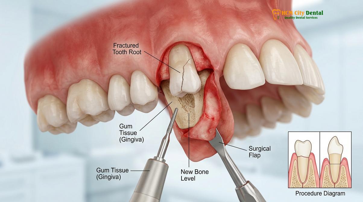
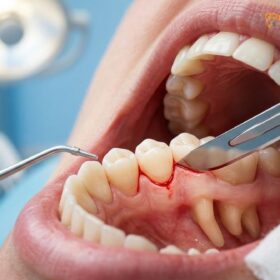
The surgical workflow for restoring these compromised teeth involves a combination of osteotomy (the removal of supporting alveolar bone) and osteoplasty (the reshaping of non-supporting bone). The clinician carefully reflects a mucoperiosteal flap to gain direct visual access to the defect. Using specialized surgical burs and micro-instruments, the bone surrounding the tooth is meticulously recontoured. The goal is to drop the bone level apically so that the deepest margin of the fracture or decay is positioned at least 3 millimeters above the newly established bone crest. This precise mathematical calculation is essential to accommodate the biological width and the restorative margin.
Clinical Case Review: Restoring a Subgingival Fracture
A patient presented to HCMC Dental Clinic in Ho Chi Minh City with a severe oblique fracture of the maxillary right premolar, extending 2mm below the gingival margin. Extraction was initially feared. However, Dr. Nguyen Van Cuong performed a localized functional crown lengthening procedure, carefully recontouring the osseous crest to expose 3mm of sound root structure. Following a 6-week tissue maturation phase, a high-strength zirconia crown was successfully bonded. The tooth was saved, restoring full masticatory function and preserving the patient’s natural alveolar ridge architecture.
For patients seeking to restore fractured teeth HCMC, this procedure represents a conservative, biologically sound alternative to tooth loss. Preserving the natural tooth root maintains the proprioceptive feedback of the periodontal ligament, which is crucial for natural chewing mechanics. It also prevents the inevitable bone resorption that occurs following an extraction, preserving the natural contours of the jawbone and the overlying facial soft tissues.
The decision to perform crown lengthening versus extraction and implant placement requires a nuanced clinical judgment. Factors such as the root length, root trunk anatomy, tooth mobility, and the strategic value of the tooth within the overall dental arch must be carefully evaluated. When the anatomical conditions are favorable, functional crown lengthening provides a highly predictable and cost-effective method to extend the lifespan of the natural dentition[2].
Securing the Biological Width to Avoid Chronic Gums Inflammation
Maintaining the biological width—a minimum of 2mm of supracrestal tissue attachment—is non-negotiable to prevent chronic inflammation and unpredictable bone resorption around new restorations.
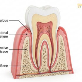
One of the most critical concepts in restorative dentistry and periodontics is the preservation of the biological width, recently reclassified in modern periodontal literature as the supracrestal tissue attachment. This anatomical dimension refers to the natural space occupied by the healthy gingival tissues above the alveolar bone crest. In a healthy state, this attachment apparatus consists of approximately 0.97 millimeters of junctional epithelium and 1.07 millimeters of connective tissue attachment, totaling roughly 2.04 millimeters. This biological seal acts as a vital barrier, protecting the underlying bone from the bacteria present in the oral cavity.
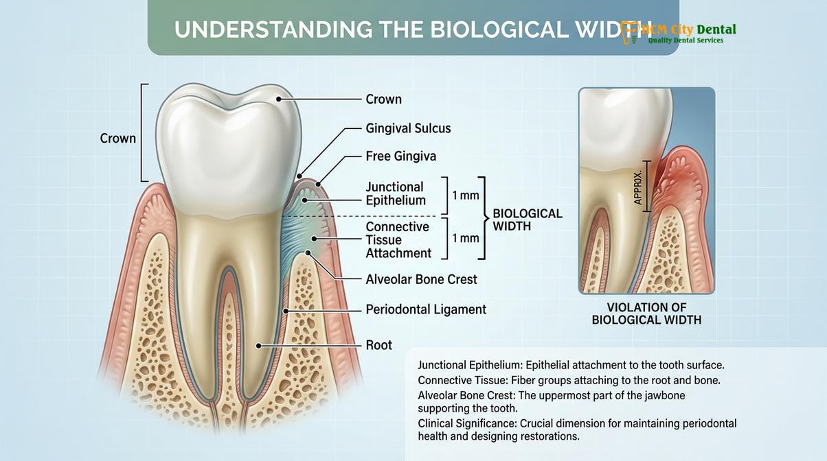
A biological width violation occurs when a dental restoration, such as a porcelain veneer or crown margin, is placed too deeply beneath the gumline, encroaching upon this sacred 2-millimeter space. When the restorative margin invades the supracrestal tissue attachment, the body perceives the restoration as a foreign object. This triggers a chronic, localized inflammatory response as the immune system attempts to recreate the necessary space between the bone and the invading margin.
Clinical Warning: The Consequences of Biological Width Violation
Placing veneer or crown margins too close to the alveolar bone without proper surgical crown lengthening will inevitably lead to chronic gingival inflammation. Patients will experience persistent bleeding upon brushing, localized swelling, a purplish discoloration of the gums, and eventual unpredictable bone resorption. This not only compromises the aesthetics of the smile but also leads to the premature failure of the dental restorations.
To prevent this cascade of pathological events, clinicians must perform meticulous biological width sounding prior to finalizing any treatment plan. According to Dr. Nguyen Van Cuong, precise calibration of the gingival zenith points and thorough bone sounding are mandatory diagnostic steps. If the distance from the proposed restorative margin to the bone crest is less than 3 millimeters (allowing 2mm for the attachment and 1mm for the sulcus), surgical crown lengthening is absolutely indicated to lower the bone level and recreate a healthy biological dimension[3].
The healing process following the re-establishment of the biological width is highly predictable, provided the surgical execution is precise. The tissues will naturally reattach to the root surface at the newly established level, forming a robust, healthy seal. This ensures that when the final porcelain veneers or crowns are placed, the surrounding gingiva will remain pink, stippled, and firmly adapted to the restorative margins, ensuring long-term aesthetic and functional success.
Combining Crown Lengthening with Porcelain Veneers and Crowns
Integrating crown lengthening with porcelain veneers ensures perfect gingival zenith points and symmetrical smile arcs, utilizing advanced laser technology for rapid, predictable healing.
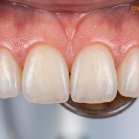
In the pursuit of the perfect smile, the white aesthetics (the teeth) and the pink aesthetics (the gums) must be in absolute harmony. Crown lengthening before veneers is frequently employed to correct discrepancies in gingival levels, treat altered passive eruption, and establish ideal tooth proportions. The golden proportion in dentistry dictates that the ideal width-to-length ratio of a maxillary central incisor should be approximately 75-80%. When teeth appear short and square due to excessive gingival display, aesthetic crown lengthening is the definitive solution to reveal the hidden beauty of the natural enamel.
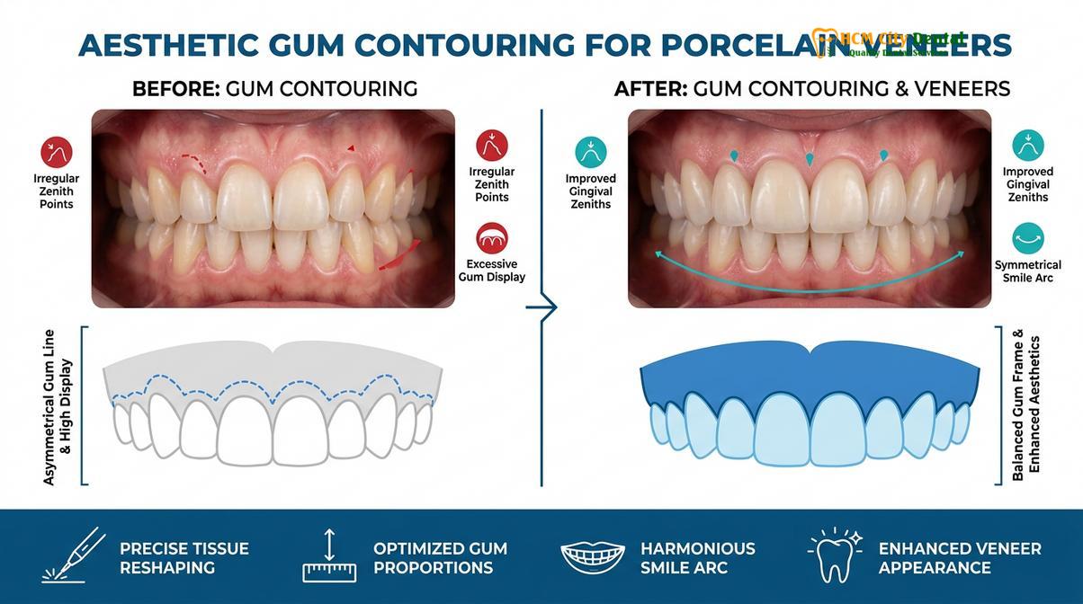
Modern aesthetic crown lengthening heavily relies on Digital Smile Design (DSD) protocols. Through high-resolution photography and digital intraoral scanning, clinicians can analyze the patient’s facial midline, interpupillary line, and lip dynamics. This digital blueprint allows for the precise calculation of the ideal gingival zenith points—the highest point of the gum scallop, which should be displaced slightly distal to the long axis of the tooth. By transferring this digital design to the surgical field via custom surgical guides, the clinician can execute the tissue contouring with micrometer accuracy.
The integration of advanced laser technology has revolutionized this procedure. At leading facilities, the use of AMD Picasso Lasers (Diode soft tissue laser, 810nm) is the gold standard for soft tissue management. The 810nm wavelength is highly absorbed by melanin and hemoglobin, making it exceptionally efficient for cutting gingival tissue while simultaneously coagulating blood vessels. This results in a virtually bloodless surgical field, providing the clinician with unparalleled visibility. Furthermore, the laser energy cauterizes nerve endings and sterilizes the surgical site, drastically reducing the risk of postoperative infection.
“The application of 810nm diode laser technology in aesthetic gingivectomy offers profound clinical advantages. It allows for sutureless incisions, instant hemostasis, and a significantly accelerated healing cascade, enabling patients to return to their normal routines with minimal discomfort.”
For international patients engaging in dental tourism, the efficiency and rapid recovery associated with laser dentistry are highly advantageous. The minimally invasive nature of the AMD Picasso Laser typically allows for a rapid 48-hour initial recovery period, with significantly reduced postoperative pain and swelling compared to traditional scalpel techniques. This accelerated timeline is crucial for patients who need to complete their comprehensive smile makeovers within a limited travel window[4].
Understanding the financial investment is an important aspect of treatment planning. The clinic maintains a transparent pricing structure for these advanced procedures. Currently, according to the latest clinic fee schedule, the costs are structured to provide exceptional value while utilizing top-tier technology.
| Procedure Type | Standard Price (Walk-in) | Online Booking Price (-40% WhatsApp) | Estimated USD |
|---|---|---|---|
| Laser Gingivectomy (per tooth) | 2,500,000 VND | 1,500,000 VND | ~$60 USD |
| Full-Arch Laser Gum Contouring (up to 10 teeth) | 20,000,000 VND | 12,000,000 VND | ~$480 USD |
| Functional/Esthetic Crown Lengthening (per tooth, incl. bone) | 5,000,000 VND | 3,000,000 VND | ~$120 USD |
| Botox Gummy Smile Correction (both sides) | 7,500,000 VND | 4,500,000 VND | ~$180 USD |
| Surgical Lip Repositioning / Lip Lowering | 25,000,000 VND | 15,000,000 VND | ~$600 USD |
Following the surgical phase, a mandatory healing period is observed before the final porcelain restorations are fabricated. While the initial soft tissue healing is rapid, the complete maturation of the gingival margins and the underlying bone can take anywhere from 6 to 12 weeks. During this phase, patients are fitted with high-quality provisional restorations that guide the healing tissue and allow the patient to “test drive” their new smile aesthetics. Only when the tissue margins are absolutely stable will the final impressions be taken for the permanent Gum Contouring & Gummy Smile restorations.
Advanced Surgical and Non-Surgical Gummy Smile Interventions
For complex gummy smiles involving hyperactive muscles or skeletal excess, treatments range from Botox injections to orthognathic surgery in collaboration with maxillofacial specialists.
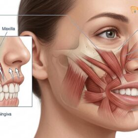
While crown lengthening addresses issues related to the teeth and gums, a gummy smile can also be caused by extra-oral factors, specifically hypermobile lips or skeletal discrepancies. A comprehensive diagnosis is essential to determine the precise etiology of the excessive gingival display. When the upper lip rises excessively upon smiling—a condition often caused by a hyperactive levator muscle—non-surgical and soft-tissue surgical interventions are highly effective.
Botox (Botulinum Toxin Type A) injections offer a minimally invasive, temporary solution for a hyperactive upper lip. By strategically injecting small units of Botox into the levator labii superioris alaeque nasi muscles, the neuromuscular transmission is temporarily blocked. This relaxes the muscles, preventing the lip from elevating excessively and effectively masking the gummy smile. The results typically last for 3 to 6 months, making it an excellent option for patients seeking a non-surgical approach or those wishing to preview the effects of a lowered lip line.
For a more permanent soft-tissue solution, surgical lip repositioning (or lip lowering surgery) is performed. This procedure involves removing a strip of mucosa from the maxillary vestibule and suturing the lip to a lower position, physically restricting its upward movement during a full smile. This technique is highly successful for patients with adequate clinical crown length but excessive lip mobility, providing a permanent enhancement to the smile dynamics.
In severe cases where the gummy smile is caused by Vertical Maxillary Excess (VME)—a skeletal condition where the upper jawbone has grown too far downward—periodontal surgery and lip repositioning alone are insufficient. These complex skeletal discrepancies require orthognathic surgery, specifically a Le Fort I osteotomy. During this major surgical procedure, the entire upper jaw is sectioned, repositioned superiorly (impacted), and stabilized with titanium plates and screws. To ensure the highest level of safety and clinical excellence for these severe skeletal cases, the clinic works in close collaboration with elite maxillofacial surgical partners at the National Hospital of Odonto-Stomatology in Ho Chi Minh City (Bệnh viện Răng Hàm Mặt Trung Ương TP.HCM), recognized as the highest clinical authority in the region[5].
When to See a Doctor for Crown Lengthening
Determining the need for crown lengthening requires a professional clinical assessment. You should schedule a consultation with a qualified dental specialist if you experience any of the following clinical signs:
- Severe Tooth Fracture: If a tooth has broken off at or below the gumline, prompt evaluation is necessary to determine if the root can be salvaged through functional lengthening.
- Deep Subgingival Decay: Cavities that extend deep beneath the gums cannot be properly filled or crowned without surgically exposing the margins.
- Excessive Gingival Display: If you feel your teeth look too short or your smile reveals an excessive amount of gum tissue, an aesthetic evaluation can determine if altered passive eruption is the cause.
- Chronic Gum Inflammation Around Existing Crowns: Persistent bleeding, swelling, or pain around older dental crowns may indicate a biological width violation that requires surgical correction.
- Preparation for Smile Makeovers: If you are planning to receive multiple porcelain veneers, an evaluation of your gingival zenith points is crucial to ensure symmetrical and harmonious results.
It is important to note that while crown lengthening has a high success rate, it is not universally applicable. Contraindications may include inadequate root length, unfavorable root anatomy, or severe systemic health issues. A personalized clinical examination, including 3D imaging and periodontal probing, is essential to formulate a safe and effective treatment plan. We encourage patients to contact HCMC Dental Clinic to schedule a comprehensive diagnostic review with our specialists.
Frequently Asked Questions
Can you place a veneer immediately after crown lengthening?
No, you generally cannot place a final veneer immediately after surgical crown lengthening. The gingival tissues require a maturation period of 6 to 12 weeks to stabilize. Placing restorations too early can result in margin exposure as the tissue shrinks during the final healing phase. During this waiting period, high-quality provisional (temporary) veneers are placed to protect the teeth, guide tissue healing, and maintain aesthetics.
What happens if you bypass functional crown lengthening?
Bypassing this crucial step often leads to a biological width violation if the restoration margin is placed too close to the bone. This results in chronic gum inflammation, localized bleeding, unpredictable bone loss, and eventual failure of the dental restoration. The body will constantly fight the invading margin, leading to a perpetual state of localized periodontitis that compromises both the tooth and the surrounding bone.
Does functional crown lengthening protect tooth roots?
Yes, functional crown lengthening helps protect the tooth root by exposing enough sound tooth structure to allow for a proper ferrule effect. This ensures the new crown or veneer fits securely, sealing the root from bacterial microleakage and preventing further subgingival decay. By establishing a solid foundation, the procedure significantly reduces the risk of root fractures under the stress of chewing.
Is laser crown lengthening painful?
Laser crown lengthening is highly tolerable and involves minimal discomfort due to the use of local anesthesia and advanced diode technology. The laser cauterizes nerve endings and blood vessels instantly, significantly reducing postoperative pain, swelling, and the need for traditional sutures. Most patients manage any mild postoperative sensitivity with over-the-counter analgesics and return to normal activities within 24 to 48 hours.
How long does the healing process take before final restorations?
Initial soft tissue healing typically occurs within 48 to 72 hours, especially when using diode lasers. However, complete osseous and gingival maturation requires approximately 6 to 12 weeks, ensuring the tissue margins are entirely stable before taking impressions for final porcelain veneers. Rushing this process can lead to aesthetic failures, as the gums may continue to recede slightly as the underlying bone remodels.
References
- Journal of Clinical Periodontology. Esthetic crown lengthening outcomes and tissue stability. (2021).
- International Journal of Periodontics & Restorative Dentistry. Diode laser vs scalpel gingivectomy in aesthetic zones. (2020).
- Journal of Aesthetic and Restorative Dentistry. Biological width around restorations and periodontal health. (2019).
- Journal of Oral and Maxillofacial Surgery. Le Fort I osteotomy stability in vertical maxillary excess. (2022).
- Clinical Oral Investigations. Surgical lip repositioning longevity and patient satisfaction. (2018).