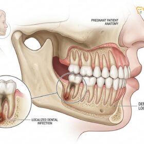
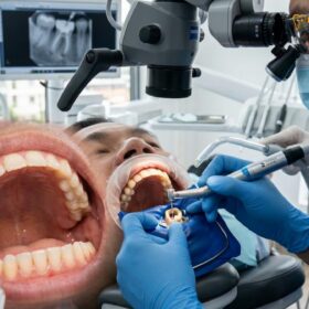
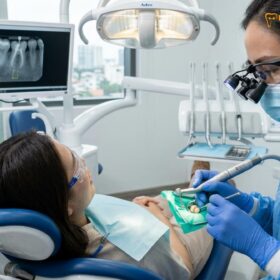
If you experience severe, throbbing tooth pain, lingering sensitivity to hot or cold, or swollen gums, these are primary need a root canal signs. These symptoms indicate that the inner pulpal tissue is inflamed or infected and requires immediate clinical intervention to save the tooth.
Clinical Summary:
Identifying the clinical indications for endodontic therapy is critical for preserving natural dentition and preventing the systemic spread of oral infections. The transition from reversible pulpitis (mild inflammation) to irreversible pulpitis and eventual pulpal necrosis is marked by specific, progressive symptoms. Patients typically first notice an exaggerated, prolonged response to thermal stimuli, which evolves into spontaneous, nocturnal pain as intrapulpal pressure increases. As the internal nerve tissue dies, the pain may temporarily subside, only to be replaced by signs of periapical infection, such as localized gingival swelling, pain upon mastication, or the formation of a draining sinus tract (fistula). Advanced diagnostic protocols, including thermal vitality testing, electric pulp testing (EPT), and Cone Beam Computed Tomography (CBCT), are utilized to confirm the extent of pulpal and periradicular disease. Timely endodontic therapy removes the necrotic debris, eradicates the bacterial biofilm, and seals the root canal system, thereby resolving the pathology and restoring the tooth to optimal function.
Key Takeaways:
- Lingering pain from hot or cold stimuli lasting more than 10-15 seconds strongly indicates irreversible pulpal damage.
- Spontaneous, throbbing pain that disrupts sleep is a hallmark of severe intrapulpal inflammation and elevated pressure.
- A single tooth turning dark gray or brown is a classic visual indicator of internal nerve death and hemorrhage.
- Localized gum swelling or a persistent pimple (fistula) signifies that a bacterial infection has spread into the surrounding jawbone.
- A sudden cessation of severe pain does not mean the tooth has healed; it often indicates complete pulpal necrosis requiring immediate intervention.
Lingering Sensitivity to Hot and Cold: The Pulpitis Indicator
Lingering pain after consuming hot or cold foods indicates irreversible pulpitis, meaning the internal nerve tissue cannot heal itself and requires professional endodontic treatment.
One of the earliest and most reliable need a root canal signs is an exaggerated and prolonged response to thermal changes. To understand why this occurs, it is necessary to examine the microscopic anatomy of the tooth. Beneath the hard, protective enamel lies the dentin, a porous layer containing thousands of microscopic channels known as dentinal tubules. These tubules radiate outward from the central pulp chamber, which houses the tooth’s vital nerves, blood vessels, and connective tissue. According to the hydrodynamic theory of tooth sensitivity, fluid within these tubules expands or contracts in response to temperature fluctuations. When the protective enamel is compromised by deep decay, a fracture, or a failing restoration, thermal stimuli cause rapid fluid movement, which directly stimulates the nerve endings within the pulp[1].
In a healthy tooth, or one with mild, reversible inflammation (reversible pulpitis), consuming a cold beverage might cause a sharp, fleeting flash of pain that dissipates within one to two seconds after the stimulus is removed. This rapid response is mediated by the A-delta nerve fibers, which are myelinated and responsible for transmitting sharp, localized pain signals. However, when the bacterial invasion penetrates deeply into the pulp chamber, the inflammatory response becomes severe and irreversible. The tissue becomes hyperemic, meaning it is engorged with blood, and the threshold for nerve activation drops significantly.
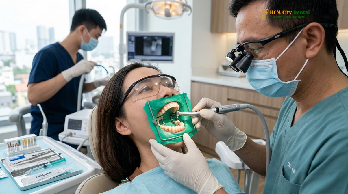
At this stage of irreversible pulpitis, the unmyelinated C-fibers within the deep pulp become heavily involved. These fibers transmit dull, aching, and poorly localized pain. When a patient with irreversible pulpitis consumes something cold—or particularly something hot—the resulting pain does not vanish immediately. Instead, they experience lingering tooth sensitivity that can last for several minutes or even hours after the thermal trigger is gone. Heat sensitivity is particularly diagnostic of advanced pulpal degeneration; as gases from necrotic tissue expand upon heating, they exert excruciating pressure on the remaining vital nerve fibers. If you find yourself avoiding hot coffee or relying on ice water to temporarily soothe a burning ache, it is a definitive signal that the pulpal tissue is failing and root canal treatment is necessary to remove the inflamed tissue.
Severe, Throbbing Toothache: When Pain Disrupts Sleep
A spontaneous, throbbing toothache that worsens at night or disrupts sleep is a classic hallmark of severe pulpal inflammation and rising intrapulpal pressure.
While mild sensitivity can sometimes be attributed to gum recession or minor enamel wear, a spontaneous, intense toothache that occurs without any external trigger is a profound medical warning. This type of pain is frequently described as a deep, pulsating ache that synchronizes with the patient’s heartbeat. The physiological mechanism behind this throbbing tooth pain is rooted in the unique anatomical constraints of the dental pulp. Unlike most tissues in the human body, which can swell freely when inflamed (such as a sprained ankle), the dental pulp is entirely encased within the rigid, unyielding walls of dentin. It is a low-compliance environment.
When bacteria from a deep cavity reach the pulp, the body’s immune system mounts a defense by increasing blood flow to the area (vasodilation) and increasing capillary permeability to allow white blood cells to reach the infection site. This inflammatory exudate causes the tissue to swell. Because the dentin walls cannot expand to accommodate this swelling, the internal pressure within the tooth spikes dramatically. This localized “compartment syndrome” compresses the delicate venules, restricting blood outflow while arterial blood continues to pump in. The resulting ischemia (lack of oxygen) and extreme pressure on the C-fibers generate an excruciating, throbbing pain[2].
“Patients often report that their toothache becomes unbearable the moment they lie down to sleep. This is a classic physiological response; moving from a standing to a supine position increases blood pressure in the head, which directly exacerbates the already critical intrapulpal pressure, making sleep virtually impossible without clinical intervention.”
— Dr. Nguyen Van Cuong, Lead Endodontist
This nocturnal exacerbation is a hallmark symptom that dentists look for during diagnosis. The pain is often so severe that over-the-counter analgesics like ibuprofen or acetaminophen provide little to no relief. Furthermore, the pain may radiate along the trigeminal nerve pathways, causing referred pain in the jaw, ear, or temple, making it difficult for the patient to pinpoint exactly which tooth is the source of the agony. Prompt endodontic intervention is required to open the tooth, release the trapped pressure, and remove the dying nerve tissue, which provides almost immediate relief from the throbbing sensation.
Swollen or Tender Gums: Signs of an Active Bacterial Abscess
Localized gum swelling near the root apex signifies that a bacterial infection has exited the tooth and formed an abscess in the surrounding jawbone.
As pulpal disease progresses untreated, the nerve tissue eventually succumbs to the infection and dies (necrosis). However, the bacteria—often a complex polymicrobial biofilm dominated by strict anaerobes—continue to multiply within the hollow root canal system. Because the tooth’s blood supply has been severed, the immune system cannot reach inside the tooth to clear the infection. Consequently, the bacteria and their toxic byproducts (such as lipopolysaccharides) migrate down the root canal and exit through the apical foramen (the opening at the tip of the root) into the surrounding periradicular tissues.
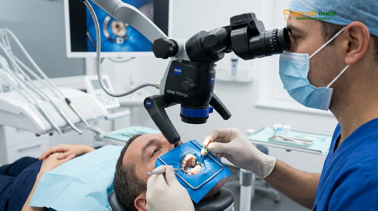
This invasion triggers a massive inflammatory response in the alveolar bone, a condition known as apical periodontitis. The immune system attempts to wall off the infection by deploying macrophages, neutrophils, and lymphocytes, which release cytokines that stimulate osteoclasts to resorb the surrounding bone. This creates a void filled with purulent exudate (pus), dead white blood cells, and necrotic debris—clinically recognized as a periapical abscess. When this abscess expands, it pushes against the cortical plate of the jawbone and the overlying gingival tissues, resulting in noticeable swelling[3].
Clinical Warning: Swelling that extends beyond the immediate gum area, causing facial asymmetry, swelling of the cheek, jaw, or neck, or accompanied by a fever and difficulty swallowing, indicates a spreading fascial space infection (such as cellulitis or Ludwig’s angina). This is a medical emergency requiring immediate clinical care and systemic antibiotics alongside dental treatment.
The swelling associated with a root canal infection can range from a small, tender nodule localized directly over the root tip to diffuse, fluctuant swelling that distorts the patient’s facial features. The gums in the affected area will typically appear highly erythematous (red), engorged, and bleed easily upon probing. Palpating the gums over the root apex will usually elicit a sharp pain response. In these scenarios, saving the infected tooth requires not only cleaning and shaping the internal canal system but also establishing proper drainage to resolve the acute abscess and allow the surrounding bone to heal.
Discoloration of the Tooth: Internal Pulpal Necrosis
A tooth that turns dark gray, brown, or black from the inside out is exhibiting signs of internal pulpal necrosis, where dying tissue stains the dentin.
While teeth naturally yellow over time due to the thinning of enamel and the deposition of secondary dentin, a sudden or localized discoloration of a single tooth is a significant diagnostic indicator. When a tooth sustains physical trauma—such as a sports injury, a fall, or a severe impact—the delicate blood vessels entering the root apex can be severed or severely damaged. This trauma can induce pulpal hemorrhage, where red blood cells rupture and spill their contents into the pulp chamber.
As these erythrocytes break down, they release hemoglobin. The iron within the hemoglobin reacts with hydrogen sulfide produced by anaerobic bacteria (if present) to form iron sulfide, a dark compound that permeates the porous dentinal tubules. Additionally, the degradation of necrotic nerve tissue produces porphyrins and other pigmented byproducts. Because dentin is highly permeable, these dark pigments stain the tooth from the inside out, causing the clinical crown to take on a dull, opaque, grayish, brown, or even black appearance. This intrinsic staining cannot be resolved with surface whitening treatments or abrasive toothpastes.
Clinical Case Example: A 28-year-old patient visited HCMC Dental Clinic in Ho Chi Minh City complaining of a front incisor that had slowly turned dark gray over two years following a minor bicycle accident. The tooth was completely asymptomatic. Clinical vitality testing revealed no response to cold or electric stimuli, confirming complete pulpal necrosis. Following a successful root canal procedure and internal bleaching, the tooth’s natural shade was restored, and the periapical bone healed seamlessly.
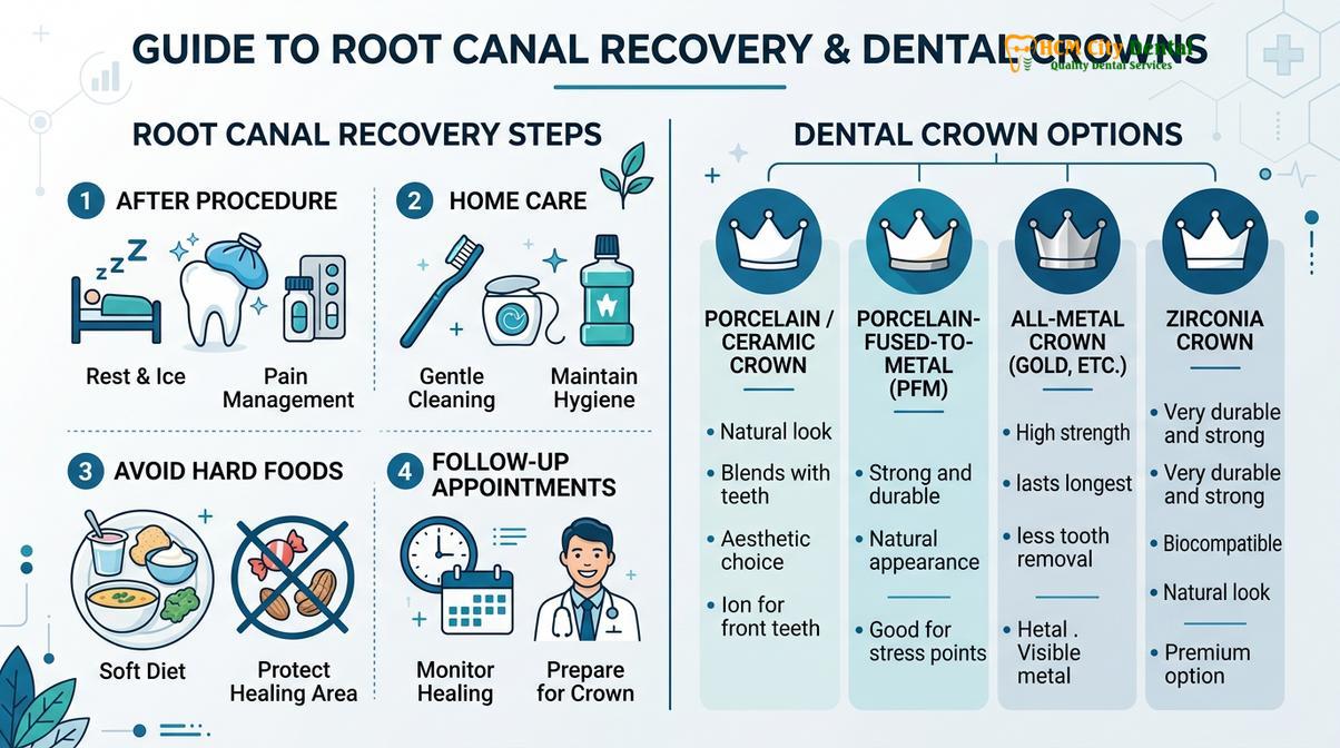
Discoloration is one of the most definitive signs of tooth nerve death. It is crucial to understand that a discolored tooth may not cause any pain, as the sensory nerves within have already perished. However, the necrotic tissue acts as a breeding ground for bacteria, which will inevitably lead to a periapical infection if left untreated. Endodontic therapy removes the stained, necrotic debris from the pulp chamber and seals the space, preventing further infection and preparing the tooth for aesthetic restoration, such as a ceramic crown or internal bleaching.
Pain when Chewing or Applying Pressure: Periodontal Membrane Inflammation
Sharp pain upon biting or applying pressure indicates that the infection has spread to the periodontal ligament, causing inflammation at the base of the root.
A healthy tooth is suspended in its bony socket by the periodontal ligament (PDL), a complex network of collagen fibers, blood vessels, and nerves. The PDL acts as a biological shock absorber, distributing the immense forces generated during mastication (chewing). It is also densely populated with proprioceptors and mechanoreceptors, which provide the brain with precise feedback regarding bite pressure and tooth position.
When a pulpal infection advances through the root apex and causes apical periodontitis, the inflammatory exudate accumulates in the confined space between the root tip and the alveolar bone. This localized edema physically pushes the tooth slightly out of its socket—often by just a fraction of a millimeter, but enough to alter the patient’s occlusion. When the patient bites down, the tooth is forced back into the inflamed, fluid-filled socket, compressing the highly sensitized nerve endings within the PDL. This results in a sharp, exquisite pain upon chewing or even light tapping (percussion) of the tooth[4].
| Clinical Condition | Nature of Pain | Diagnostic Indicators |
|---|---|---|
| Apical Periodontitis (Needs Root Canal) | Dull to sharp pain upon any pressure; tooth feels “high” in the bite. | Negative vitality tests; pain on vertical percussion; periapical radiolucency on X-ray. |
| Cracked Tooth Syndrome | Sharp, shooting pain specifically upon releasing the bite. | Positive vitality tests; pain localized to specific cusps; visible fracture lines under magnification. |
| Periodontal Abscess | Dull ache; pain on lateral pressure. | Deep periodontal pockets; vital pulp; swelling higher up on the gingival margin. |
During a clinical examination, dentists utilize a percussion test—gently tapping the biting surface of the tooth with the handle of a dental mirror—to evaluate the status of the PDL. A highly positive percussion response is a strong indicator that the infection has breached the confines of the root canal system. Professional root canal therapy addresses this by thoroughly disinfecting the internal canal space, which removes the source of the bacterial toxins. Once the root canal is sealed, the immune system can clear the periapical inflammation, allowing the PDL to heal and the pain upon chewing to subside completely.
A Persistent Pimple on the Gums: The Fistula Drainage Path
A recurring bump on the gums, known as a dental fistula or parulis, is a drainage tract for pus originating from a chronic root canal infection.
In cases of chronic apical abscesses, the body attempts to manage the continuous production of purulent exudate (pus) by creating a pressure-relief valve. The pus, which consists of dead bacteria, white blood cells, and tissue debris, follows the path of least resistance through the alveolar bone. It slowly erodes the cancellous bone, perforates the outer cortical plate, and tunnels through the soft gingival tissue, eventually forming a small, pimple-like bump on the surface of the gums. This anatomical pathway is medically termed a sinus tract, and the visible bump is called a parulis or dental fistula.
Patients often report that this gum pimple fluctuates in size, occasionally rupturing to release a foul-tasting, salty fluid into the mouth, which may also cause chronic halitosis (bad breath). The rupture of the fistula temporarily relieves the pressure within the jawbone, which is why teeth with active sinus tracts are frequently asymptomatic or cause only mild discomfort. However, the absence of severe pain is highly deceptive. The continuous drainage indicates a robust, ongoing bacterial infection that is actively destroying the surrounding jawbone.
Attempting to “pop” or treat the gum pimple with topical antiseptics is entirely ineffective, as the source of the infection lies deep within the necrotic root canal system. To accurately diagnose the origin of the fistula, an endodontist may perform a procedure called fistulography, where a flexible gutta-percha cone is gently threaded into the sinus tract before taking a radiograph. The cone acts as a pointer, leading directly to the infected root apex. The only definitive treatment is to eliminate the bacterial reservoir inside the tooth via endodontic therapy, after which the sinus tract will naturally close and heal within a few days.
Silent Nerve Death: Why a Chronic Infection Can Stop Hurting Temporarily
A sudden cessation of severe tooth pain does not mean the infection is cured; it often indicates complete nerve death, allowing the infection to spread silently.
One of the most dangerous misconceptions in dental health is the belief that if a toothache goes away on its own, the tooth has healed. In reality, the dental pulp has a very limited capacity for self-repair once irreversible inflammation sets in. If a patient endures days of severe, throbbing pain that suddenly vanishes, it is rarely a sign of recovery. Instead, it typically signifies that the inflammatory pressure has completely strangulated the blood supply, leading to total pulpal necrosis. The sensory nerves have died, rendering the tooth incapable of transmitting pain signals to the brain.
“We frequently see patients who delayed treatment because their severe toothache miraculously stopped. Unfortunately, this ‘silent phase’ is when the necrotic bacteria multiply unchecked, migrating into the jawbone to form large cysts or abscesses that complicate the eventual treatment.”
— Dr. Nguyen Van Cuong, Lead Endodontist
This condition is known as asymptomatic apical periodontitis. The tooth will not respond to cold or hot stimuli, and the patient may chew normally for months or even years without discomfort. However, the necrotic debris and complex bacterial biofilms—often harboring resilient species like Enterococcus faecalis—remain entrenched within the microscopic anatomy of the root canal system[5]. Over time, these bacteria continuously release toxins into the periapical tissues, causing silent, progressive bone resorption that is only visible on radiographic imaging.
Eventually, the delicate balance between the host immune system and the bacterial load will tip, leading to an acute exacerbation known as a “phoenix abscess.” This results in a rapid onset of severe swelling, intense pain, and potential systemic complications. Therefore, recognizing the early need a root canal signs and seeking prompt clinical endodontic intervention is paramount, even if the acute pain has temporarily subsided.
When to See a Doctor (Clinical Considerations)
Determining whether a tooth requires endodontic therapy cannot be done based on symptoms alone; it requires a comprehensive clinical and radiographic evaluation by a qualified dental professional. If you experience any of the symptoms discussed—particularly lingering thermal sensitivity, spontaneous throbbing pain, or localized swelling—you should schedule an examination immediately. Delaying care not only prolongs discomfort but also decreases the long-term prognosis of the tooth and increases the complexity of the required treatment.
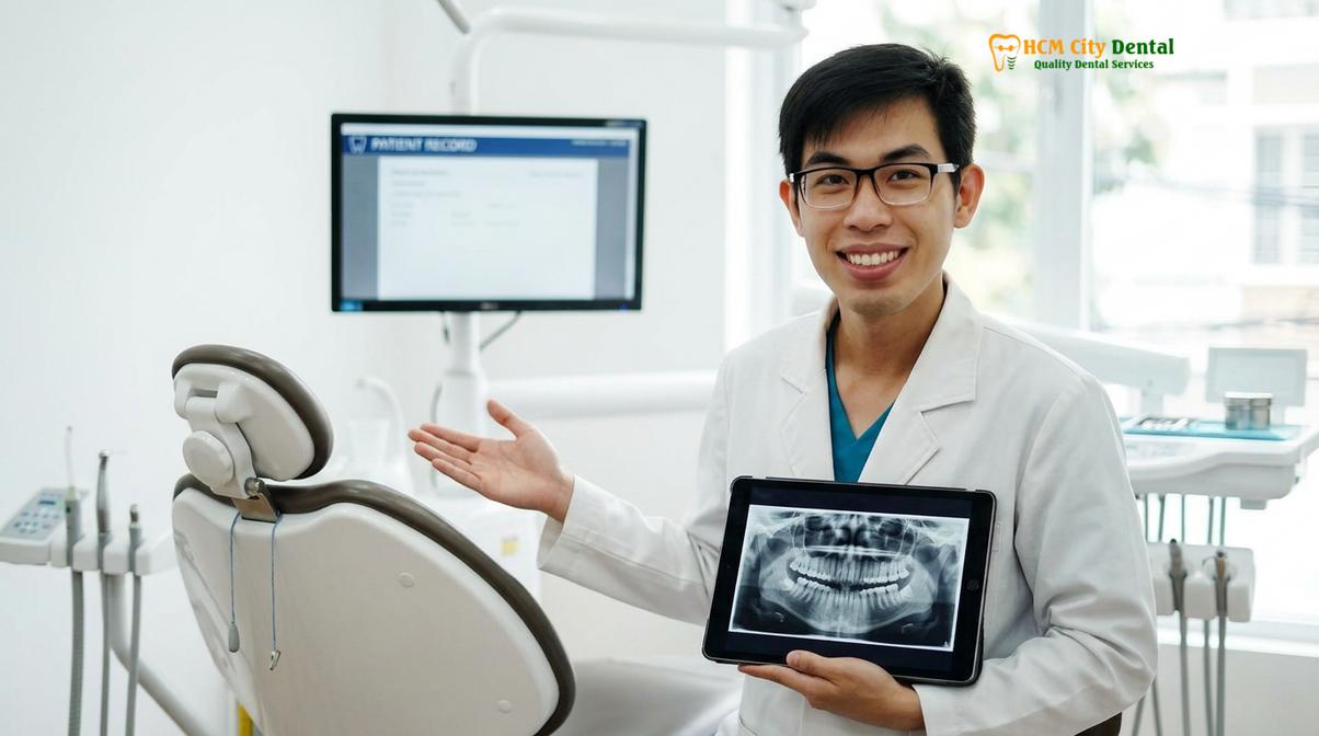
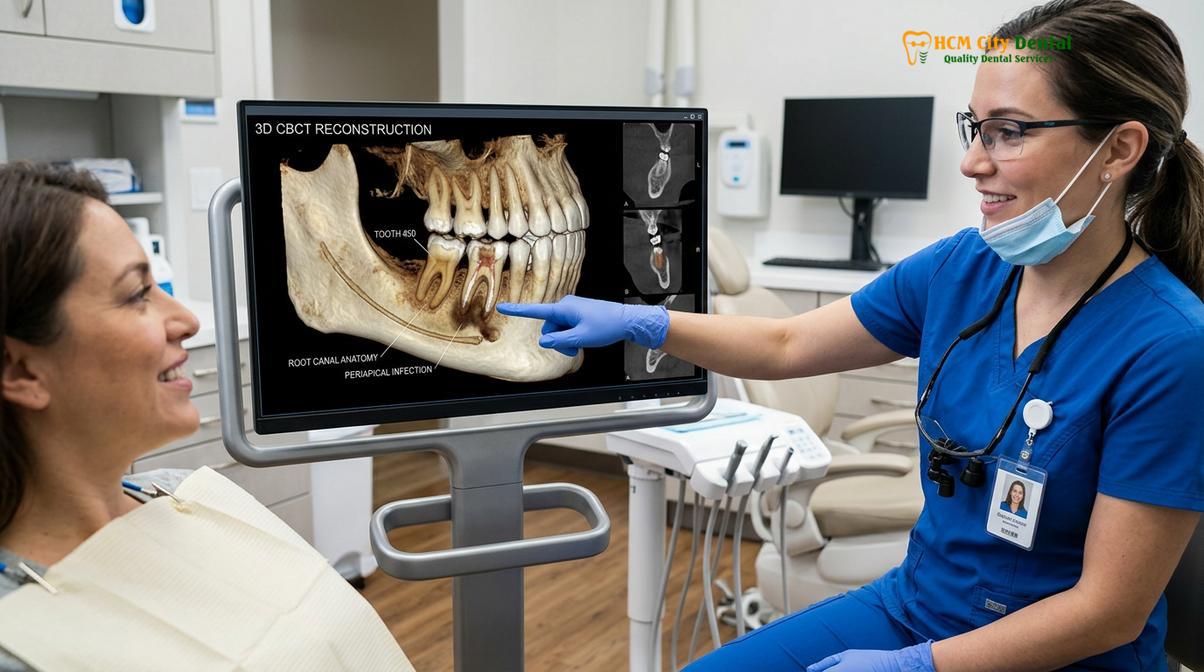
At HCMC Dental Clinic in Ho Chi Minh City, our specialists utilize advanced diagnostic tools to accurately assess pulpal health. This includes thermal vitality testing (using refrigerants like Endo-Ice), electric pulp testing (EPT) to measure nerve responsiveness, and high-resolution 3D Cone Beam Computed Tomography (CBCT) to visualize the complex root canal anatomy and detect early signs of periapical bone loss.
Red Flag Symptoms Requiring Immediate Emergency Care:
- Rapidly spreading facial swelling that affects the eye, cheek, or neck.
- Difficulty swallowing, breathing, or fully opening the mouth (trismus).
- A high fever accompanied by chills and lethargy.
- Bilateral swelling of the lower jaw.
These systemic signs indicate that the localized dental infection has breached the anatomical boundaries of the jaw and is spreading through the fascial spaces. This is a severe medical condition that requires urgent intervention, often involving intravenous antibiotics and surgical drainage in conjunction with endodontic care. For personalized diagnostics and to discuss your treatment options, a professional consultation is essential to ensure the highest standard of care and predictable clinical outcomes.
Frequently Asked Questions
Can a tooth that doesn’t hurt need a root canal?
Yes, a painless tooth can still require endodontic therapy if the internal nerve has completely died. Once pulpal necrosis occurs, the sensory nerves no longer transmit pain signals, but the bacterial infection remains active within the root canal system and can silently destroy the surrounding jawbone. Routine radiographic examinations often detect these asymptomatic infections before they flare up into painful abscesses.
How do I know if my toothache is just a cavity?
A standard cavity typically causes mild, fleeting sensitivity to sweets or cold that disappears immediately after the stimulus is removed. In contrast, if the pain is severe, throbbing, wakes you up at night, or lingers for minutes after drinking cold water, the decay has likely reached the pulp, necessitating endodontic intervention. A clinical vitality test is required to confirm the exact depth of the damage.
Will antibiotics cure a root canal infection without a dentist?
No, antibiotics cannot cure an infected root canal because the blood supply to the dead nerve is severed. Without blood flow, systemic antibiotics cannot reach the bacteria hiding inside the hollow tooth structure; the infection must be physically removed through professional chemo-mechanical debridement. Antibiotics are only prescribed as an adjunct to manage spreading swelling or systemic symptoms.
Does a dark tooth always require endodontic therapy?
A single tooth that turns dark gray or black usually indicates internal bleeding or nerve death, which strongly suggests the need for endodontic treatment. However, a comprehensive clinical examination, including vitality testing and radiographic imaging, is required to confirm the diagnosis before initiating therapy. In some rare cases of calcific metamorphosis, the tooth may discolor but remain vital and asymptomatic.
How long can I delay treatment if I see a gum pimple?
You should not delay treatment if you notice a pimple on your gums, as this indicates an active, draining bacterial abscess. Postponing therapy allows the infection to continuously erode the surrounding alveolar bone, potentially compromising adjacent teeth and increasing the risk of systemic bacterial spread. Prompt treatment ensures the highest success rate for saving the natural tooth.
References
- Journal of Endodontics. Outcomes of root canal treatment and restoration longevity. (2020).
- International Endodontic Journal. Biofilms in endodontics and disinfection strategies. (2021).
- Journal of the American Dental Association. Microscope-assisted endodontics and missed canals. (2019).
- Clinical Oral Investigations. Biocompatibility of bioceramic root canal sealers. (2022).
- British Dental Journal. Pain management and dental anxiety in endodontics. (2018).