Deep cleaning recovery typically takes five to seven days, with initial soreness and mild bleeding subsiding within the first 48 hours. Complete gingival reattachment to the tooth roots requires several weeks. Adhering to a soft diet and modified oral hygiene ensures optimal healing and prevents post-procedural infection.
Clinical Summary:
Recovery from periodontal scaling and root planing (SRP) involves a predictable biological timeline governed by the body’s natural inflammatory and regenerative processes. Patients generally experience transient dentinal hypersensitivity, localized gingival inflammation, and minor spotting for 24 to 48 hours following the intervention. The primary objective during the deep cleaning recovery phase is to facilitate the reattachment of the gingival tissues to the newly smoothed root surfaces, thereby reducing periodontal pocket depths. Clinical management focuses heavily on biofilm control, mechanical trauma avoidance, and thermal sensitivity mitigation. Dr. Nguyen Van Cuong emphasizes that strict adherence to post-operative protocols—including a modified soft diet, specialized antimicrobial rinses, and gentle brushing techniques—significantly accelerates tissue regeneration and minimizes the risk of secondary periodontal infections for patients treated at HCMC Dental Clinic.
Key Takeaways:
- Initial gum tenderness and mild bleeding typically resolve within 24 to 48 hours post-procedure.
- Thermal sensitivity is a normal physiological response due to temporary dentinal tubule exposure.
- A soft, nutrient-rich diet is mandatory for the first 72 hours to prevent mechanical trauma to healing gums.
- Aggressive brushing must be paused; use ultra-soft bristles and prescribed antimicrobial rinses.
- Complete gingival reattachment and pocket depth reduction take approximately four to six weeks.
What to Expect Immediately After Deep Cleaning
Immediately following your procedure, expect localized numbness, mild gingival swelling, and slight blood-tinged saliva as the local anesthesia wears off and the initial inflammatory healing cascade begins.
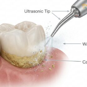
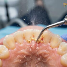
The immediate aftermath of a scaling and root planing procedure is characterized by a series of distinct physiological responses. Because the treatment involves meticulous instrumentation below the gumline to remove hardened calculus and bacterial biofilm, the periodontal tissues undergo a controlled level of mechanical trauma. This is a necessary step to halt the progression of periodontal disease, but it initiates an acute inflammatory response. As the local anesthesia begins to dissipate—typically within two to four hours—patients will start to regain sensation in their lips, cheeks, and gingival tissues. It is during this transition that the initial signs of recovery manifest.
One of the most common immediate experiences is localized tenderness and a throbbing sensation in the gums. This occurs because the blood vessels in the gingival tissues dilate to bring immune cells and healing factors to the treated areas. The removal of subgingival calculus leaves behind a raw, microscopic wound surface inside the periodontal pocket. The body immediately begins to form a delicate fibrin clot within this space, which serves as the foundational matrix for future tissue reattachment. Consequently, patients may notice pink-tinged saliva or minor oozing from the gums. This slight bleeding is a normal part of the hemostatic process and should not be a cause for alarm unless it becomes heavy or continuous.
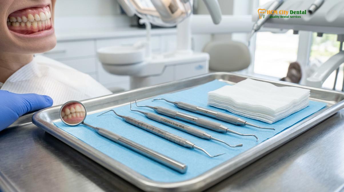
Managing pain after deep cleaning teeth requires an understanding of the inflammatory cascade. The mechanical debridement stimulates the release of prostaglandins, which are lipid compounds that mediate inflammation and pain. To counteract this, dental professionals often recommend taking over-the-counter non-steroidal anti-inflammatory drugs (NSAIDs), such as ibuprofen, before the anesthesia completely wears off. This preemptive pharmacological approach helps to blunt the peak of the inflammatory response, significantly reducing post-operative discomfort. Furthermore, the application of an ice pack to the outside of the face in 15-minute intervals can induce localized vasoconstriction, thereby minimizing edema (swelling) and providing a numbing effect to the surrounding tissues [1].
Clinical Warning: Do not attempt to eat, chew gum, or drink hot liquids while your mouth is still numb from local anesthesia. The lack of sensation severely increases the risk of accidental, traumatic biting injuries to your tongue, lips, and inner cheeks, as well as the potential for thermal burns from hot beverages.
It is also crucial to understand the state of the tooth roots immediately following the procedure. The root planing aspect of the treatment involves smoothing the cementum (the outer layer of the tooth root) to remove bacterial toxins and create a biologically acceptable surface for the gums to reattach. This process temporarily exposes the microscopic dentinal tubules. As a result, as soon as the numbing subsides, you may experience a sharp, sudden sensitivity when inhaling cold air or consuming cold liquids. This is a well-documented physiological reaction and is considered a standard part of the initial recovery phase.
Healing Timeline: Day 1 to Week 2
The healing timeline progresses from acute inflammation and clot formation in the first 48 hours to gradual tissue reattachment, reduction of edema, and resolution of sensitivity by the end of the second week.
Understanding the biological progression of scaling and root planing recovery is essential for setting realistic expectations and ensuring compliance with post-operative care. The healing of periodontal tissues is a complex, highly orchestrated event that occurs in distinct phases. While the superficial symptoms may resolve quickly, the underlying cellular regeneration requires a more extended period. The timeline can be broadly categorized into the acute inflammatory phase, the proliferative phase, and the maturation phase.
During the first 24 to 48 hours (Day 1 to Day 2), the body is primarily focused on hemostasis and acute inflammation. The delicate fibrin clots that formed immediately after the procedure are stabilizing within the periodontal pockets. Macrophages and neutrophils—specialized white blood cells—migrate to the site to phagocytize (engulf and destroy) any remaining bacteria and necrotic tissue debris. Clinically, this translates to the peak period of gingival tenderness, mild swelling, and potential spotting when brushing. The gums may appear slightly redder than usual, which is a direct result of increased vascularity. It is imperative during this window to avoid any mechanical disruption of the gums, as dislodging the fibrin clot can delay healing and increase the risk of infection.
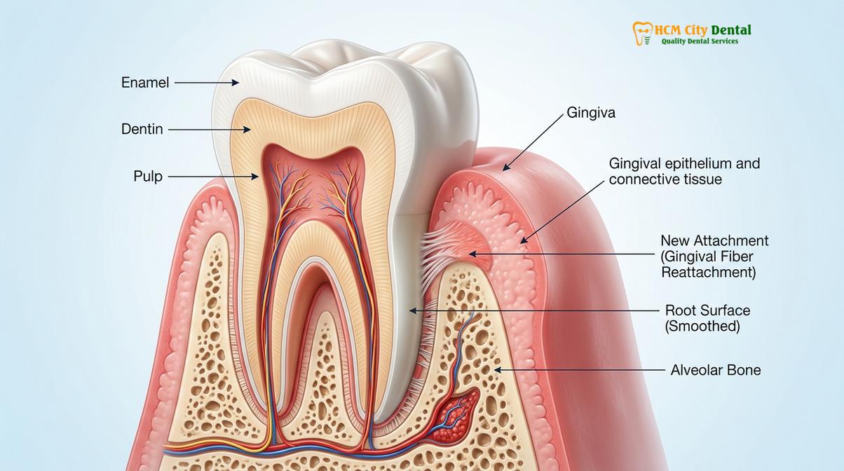
As the recovery enters Day 3 through Day 7, the acute inflammation begins to subside, marking the transition into the proliferative phase. Fibroblasts, the cells responsible for synthesizing collagen, begin to migrate into the healing area. They lay down a new extracellular matrix, which is the structural framework required for the gums to tighten around the teeth. Patients will typically notice a significant reduction in soreness and bleeding during this week. The gingival tissues will start to lose their angry red appearance, transitioning back to a healthier, stippled pink. However, thermal sensitivity may still be present, as the dentinal tubules remain partially exposed while the tissue continues to adapt to the root surface.
| Recovery Phase | Biological Process | Typical Patient Symptoms | Clinical Focus |
|---|---|---|---|
| Days 1 – 2 | Hemostasis, fibrin clot formation, acute inflammatory response. | Mild to moderate soreness, localized swelling, pink-tinged saliva, thermal sensitivity. | Pain management, avoiding mechanical trauma, maintaining the blood clot. |
| Days 3 – 7 | Fibroblast proliferation, collagen synthesis, reduction of vascular dilation. | Decreasing tenderness, cessation of bleeding, persistent but improving sensitivity. | Gentle biofilm removal, soft diet adherence, antimicrobial rinsing. |
| Week 2 | Epithelial downgrowth, formation of the long junctional epithelium. | Gums feel normal, significant reduction in sensitivity, tissue appears firm and pink. | Resuming normal (but gentle) oral hygiene, monitoring for any signs of delayed healing. |
| Weeks 4 – 6 | Connective tissue maturation, maximum pocket depth reduction achieved. | Complete resolution of symptoms, improved bite stability. | Clinical re-evaluation, periodontal probing to assess treatment success. |
By the second week of recovery, the epithelial cells have rapidly multiplied and migrated down the inner wall of the gingival sulcus, forming what is known as the long junctional epithelium. This creates a new, tighter seal against the tooth root, effectively reducing the depth of the periodontal pockets that necessitated the periodontal therapy protocols in the first place. For most patients, the gums will feel entirely normal by this point, and the hypersensitivity will have largely resolved as the tubules become naturally occluded by minerals from saliva and toothpaste. It is crucial to maintain excellent, albeit gentle, oral hygiene during this phase to prevent the recolonization of pathogenic bacteria, which could disrupt the final stages of tissue maturation.
Managing Post-SRP Sensitivity and Soreness
Post-procedural sensitivity is managed through the use of desensitizing toothpastes, avoiding extreme temperatures, and utilizing over-the-counter anti-inflammatory medications to block pain pathways.
One of the most frequently reported sequelae following periodontal debridement is dentinal hypersensitivity. To effectively manage this symptom, it is helpful to understand the underlying mechanism, known in dentistry as Brännström’s hydrodynamic theory. The roots of your teeth are covered by a thin layer of mineralized tissue called cementum, which protects the underlying dentin. Dentin is highly porous, containing thousands of microscopic channels (dentinal tubules) that run directly to the dental pulp, where the tooth’s nerve resides. During a deep cleaning, the removal of tenacious calculus often results in the incidental removal of a microscopic layer of this cementum, leaving the dentinal tubules temporarily exposed to the oral environment.
When these exposed tubules encounter stimuli—such as a cold beverage, hot soup, or even a sharp intake of cold air—the fluid inside the tubules expands or contracts rapidly. This sudden fluid movement exerts pressure on the mechanoreceptors of the A-delta nerve fibers within the pulp, translating into a sharp, transient, and often intense pain. This sensitivity after SRP is a physiological reality of the healing process, not an indication that the procedure was performed incorrectly or that the tooth is damaged [2].
“Post-scaling sensitivity is a predictable biological response, not a complication. The key to managing it lies in occluding the exposed dentinal tubules and calming the pulpal nerve. We strongly advise our patients to transition to a potassium nitrate-based toothpaste immediately following their procedure, as it effectively depolarizes the nerve endings and provides significant relief within a few days of consistent use.”
— Dr. Nguyen Van Cuong
Pharmacological management is the first line of defense against both soreness and sensitivity. Over-the-counter NSAIDs, such as ibuprofen or naproxen sodium, are highly effective because they inhibit the cyclooxygenase (COX) enzymes, thereby reducing the synthesis of prostaglandins that cause inflammation and lower the pain threshold. For patients who cannot tolerate NSAIDs due to gastrointestinal issues or other medical contraindications, acetaminophen is a viable alternative for pain relief, though it lacks the potent anti-inflammatory properties of NSAIDs. It is generally recommended to take these medications on a scheduled basis for the first 48 hours, rather than waiting for the pain to become severe.
Topical management is equally critical. Desensitizing toothpastes containing active ingredients like potassium nitrate or stannous fluoride should be incorporated into your daily routine. Potassium nitrate works by penetrating the dentinal tubules and preventing the nerve from repolarizing, effectively blocking the transmission of the pain signal. Stannous fluoride, on the other hand, works mechanically by precipitating minerals that physically plug or occlude the open tubules, stopping the fluid movement altogether. For maximum efficacy, patients are often advised to apply a small amount of desensitizing toothpaste directly to the sensitive areas with a clean finger before bed, leaving it on overnight without rinsing. Avoiding highly acidic foods and beverages (like citrus juices and sodas) is also vital, as acid can dissolve the smear layer and further open the tubules, exacerbating the sensitivity.
Diet Guide: What to Eat and Avoid After Deep Cleaning
For the first few days, consume a soft, lukewarm diet rich in proteins and vitamins while strictly avoiding crunchy, spicy, acidic, or extreme-temperature foods that can irritate healing gums.
Nutrition plays a profound role in wound healing, and the recovery period following a deep cleaning is no exception. The periodontal tissues require specific macro and micronutrients to synthesize collagen, rebuild the extracellular matrix, and mount an effective immune response against oral bacteria. However, the physical consistency and temperature of the food you consume are just as important as its nutritional profile. The primary goal of the post-SRP diet is to provide necessary sustenance while absolutely minimizing mechanical trauma and chemical irritation to the fragile, healing gingival sulcus.
When considering what to eat after deep cleaning, the focus should be on soft, nutrient-dense foods that require minimal mastication (chewing). For the first 48 to 72 hours, your diet should consist primarily of items that can be easily mashed with a fork or swallowed with little effort. Excellent choices include lukewarm broths and blended soups, which provide hydration and essential minerals without thermal shock. Scrambled eggs are highly recommended as they are soft and packed with high-quality protein and amino acids necessary for tissue repair. Mashed potatoes, sweet potatoes, and steamed, pureed vegetables offer complex carbohydrates for energy. Greek yogurt and cottage cheese are also beneficial, providing calcium and probiotics, provided they are consumed at a moderate temperature rather than straight from the refrigerator [3].
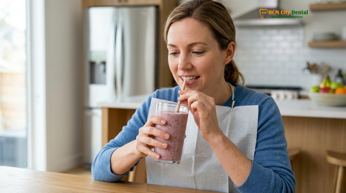
Equally critical is knowing which foods to strictly avoid. Mechanical irritants pose the greatest risk to the healing blood clot and the reattaching tissues. Hard, crunchy foods such as tortilla chips, popcorn, pretzels, and crusty breads can easily splinter into sharp fragments. If a sharp piece of food is driven into the healing periodontal pocket, it can lacerate the tissue, dislodge the fibrin clot, and introduce bacteria deep into the wound, potentially leading to a painful periodontal abscess. Similarly, foods with small, hard seeds—like strawberries, sesame seeds, and poppy seeds—should be avoided, as these tiny particles can easily become lodged beneath the gumline, causing a localized foreign body reaction and severe inflammation.
Chemical and thermal irritants must also be eliminated from the diet during the initial recovery phase. Highly acidic foods and beverages, including tomatoes, citrus fruits, vinegar-based dressings, and carbonated sodas, can cause a severe burning sensation on the raw gingival tissues and exacerbate dentinal hypersensitivity by opening the dentinal tubules. Spicy foods containing capsaicin will similarly irritate the healing wounds and increase localized blood flow, potentially triggering secondary bleeding. Finally, extreme temperatures should be avoided. Piping hot coffees or teas can dissolve the stabilizing blood clot and cause thermal burns to the anesthetized or sensitive tissues, while ice-cold beverages will trigger the sharp, shooting pain associated with exposed root surfaces. Sticking to a lukewarm, bland, and soft diet ensures that the biological healing process proceeds without unnecessary interruption.
Daily Oral Hygiene Adjustments During Recovery
Modify your daily routine by using an ultra-soft toothbrush, avoiding aggressive flossing in treated areas, and incorporating warm saltwater or prescribed chlorhexidine rinses to maintain a low-bacteria environment.
Maintaining oral hygiene after a professional dental cleaning presents a clinical paradox: the treated areas must be kept meticulously clean to prevent bacterial recolonization, yet the tissues are too fragile to withstand standard brushing and flossing techniques. Navigating this delicate balance requires specific modifications to your daily routine. The objective is to achieve effective biofilm disruption without causing mechanical trauma to the newly forming epithelial attachment. Failure to adapt your hygiene practices can result in delayed healing, increased recession, or the recurrence of periodontal inflammation.
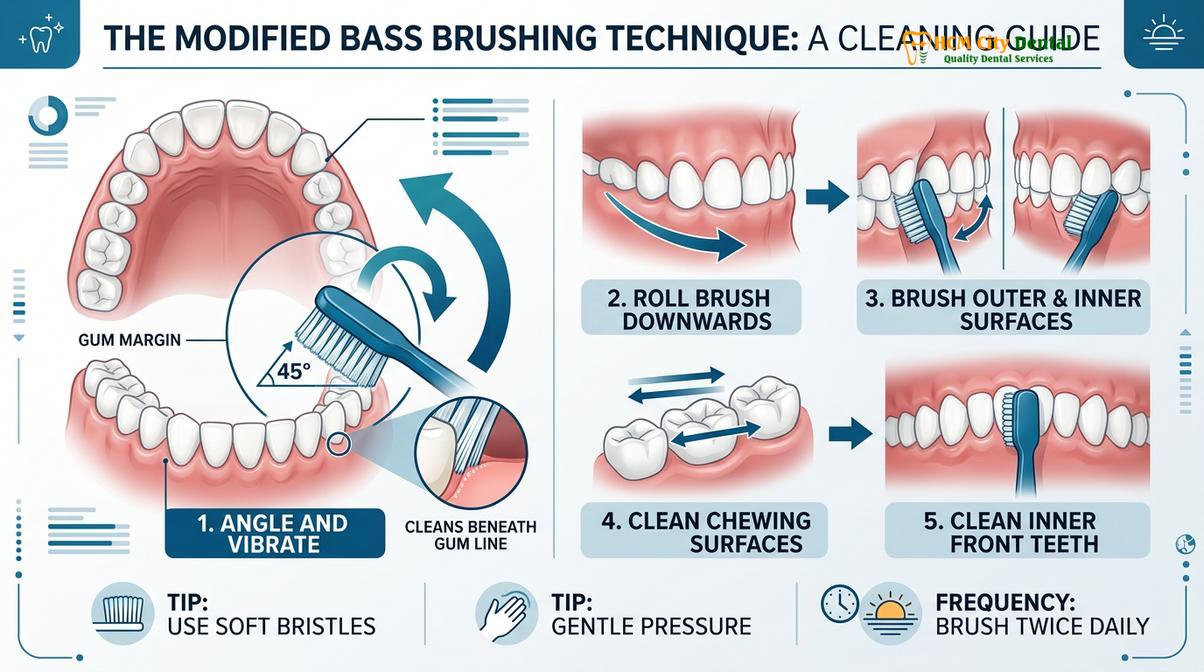
The first and most crucial adjustment is the transition to an ultra-soft bristled toothbrush. Standard medium or hard bristles are strictly contraindicated, as they will lacerate the healing gingiva and abrade the exposed root surfaces. When brushing, the technique must be modified. Dental professionals recommend a gentle variation of the Bass technique. Place the ultra-soft bristles at a 45-degree angle to the gumline and use micro-vibratory, circular motions rather than aggressive horizontal scrubbing. The goal is to gently sweep away soft plaque without forcing the bristles deep into the healing sulcus. If certain areas are too tender to brush even with an ultra-soft brush, it is acceptable to carefully wipe the teeth with a clean, damp piece of gauze for the first 24 hours, gradually reintroducing the brush as tolerance improves.
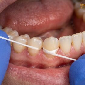
Flossing requires extreme caution during the initial recovery phase. Aggressive snapping of dental floss between the teeth can easily slice through the delicate long junctional epithelium that is attempting to reattach to the tooth root. For the first 48 to 72 hours, it is generally advised to avoid flossing deeply into the gumline in the specifically treated areas. When you do resume interdental cleaning, use a gentle, sliding motion, hugging the side of the tooth (creating a C-shape with the floss) and carefully moving it up and down without forcing it beneath the gingival margin. Water flossers can be an excellent adjunct, but they must be set to the absolute lowest pressure setting to avoid blasting apart the healing tissue matrix [4].
Clinical Case Review: A 45-year-old patient presented to HCMC Dental Clinic in Ho Chi Minh City with moderate periodontitis requiring comprehensive scaling and root planing. Post-operatively, the patient was instructed to pause standard flossing and utilize a 0.12% Chlorhexidine gluconate rinse twice daily, alongside ultra-soft brushing. By the two-week follow-up, clinical evaluation revealed a remarkable reduction in gingival erythema (redness) and a significant decrease in pocket depths, demonstrating the efficacy of chemical plaque control when mechanical cleaning must be minimized to protect tissue reattachment.
Chemical plaque control becomes an indispensable tool when mechanical cleaning is restricted. Warm saltwater rinses (one-half teaspoon of salt dissolved in eight ounces of warm water) are highly recommended starting 24 hours after the procedure. The hypertonic saline solution helps to draw out localized edema (swelling) through osmosis, alters the pH of the oral cavity to inhibit bacterial growth, and provides a soothing effect to the raw tissues. In cases of more advanced periodontal disease, your dentist may prescribe an antimicrobial mouthwash containing Chlorhexidine gluconate. This powerful antiseptic binds to the oral tissues and slowly releases over time (a property known as substantivity), effectively rupturing the cell membranes of pathogenic bacteria and maintaining a sterile environment conducive to rapid healing.
“The success of comprehensive gum disease treatment relies heavily on the patient’s home care during the recovery window. We instruct our patients to view their oral hygiene not as a vigorous scrubbing task, but as a gentle, therapeutic process. Utilizing chemical adjuncts like warm saline allows the tissue to heal undisturbed while keeping the bacterial load below the threshold of infection.”
— Dr. Nguyen Van Cuong
When to Contact Your Dentist After SRP
While the vast majority of scaling and root planing procedures heal uneventfully, it is critical to differentiate between normal physiological recovery and the signs of a developing complication. The inflammatory response—characterized by mild redness, slight swelling, and transient tenderness—is a necessary precursor to healing. However, when these symptoms escalate rather than subside, clinical intervention may be required to prevent localized tissue destruction or systemic involvement.
Bleeding is a primary indicator of healing status. As previously noted, slight pink-tinged saliva or minor oozing is expected for the first 24 to 48 hours. However, if you experience frank, bright red bleeding that fills the mouth rapidly, or bleeding that persists continuously beyond the second day despite applying gentle pressure with damp gauze, you must contact your dental provider immediately. This could indicate an unmanaged localized hemorrhage or a disruption of the vascular network that requires professional hemostatic control.
Infection is the most significant risk during the recovery phase. The oral cavity is densely populated with bacteria, and the open periodontal pockets provide a direct portal of entry. Signs of a post-operative infection include a sudden, severe increase in pain that is unresponsive to over-the-counter analgesics, progressive swelling that extends beyond the gums into the cheek or jaw, and the presence of purulent exudate (pus) discharging from the gumline. Systemic symptoms, such as a fever, chills, or swollen lymph nodes in the neck and submandibular region, are definitive red flags indicating that the localized infection has spread. If any of these symptoms manifest, prompt evaluation and the potential administration of systemic antibiotics are necessary to manage the infection and protect the integrity of the routine scaling procedures and overall periodontal health [5].
Finally, persistent hypersensitivity that does not improve after several weeks of using desensitizing protocols should be evaluated. While sensitivity is normal initially, a sharp, lingering pain that wakes you up at night or occurs spontaneously without a thermal trigger may indicate that the pulpal tissue has become irreversibly inflamed, necessitating further endodontic diagnostics. Regular follow-up appointments are a cornerstone of advanced periodontal care, ensuring that the healing trajectory is on course and that the foundational health of your smile is fully restored.
Frequently Asked Questions
How long does soreness last after deep cleaning?
Soreness typically lasts for 24 to 48 hours following a deep cleaning procedure. During this initial phase, the gingival tissues are recovering from mechanical instrumentation and localized inflammation. Over-the-counter analgesics, such as ibuprofen, and warm saltwater rinses can effectively manage this transient discomfort. If severe, throbbing pain persists beyond a few days or worsens, a clinical evaluation is recommended to rule out potential complications like a localized infection.
Can I eat immediately after scaling and root planing?
You should wait until the local anesthesia has completely worn off before eating, which usually takes two to four hours. Eating while numb poses a severe risk of accidental biting injuries to your tongue, lips, or inner cheeks, which can cause significant trauma. Once sensation fully returns, begin with a soft, lukewarm diet to avoid irritating the healing periodontal tissues and prevent mechanical disruption of the blood clot.
Why are my teeth so sensitive to cold after deep cleaning?
Teeth become sensitive to cold because the procedure removes insulating calculus and a microscopic layer of cementum, exposing the underlying dentinal tubules. According to the hydrodynamic theory, cold temperatures cause fluid movement within these microscopic tubules, which stimulates the nerve endings inside the tooth pulp. This sensitivity is usually temporary and gradually subsides as the tissues heal, remineralize, and respond to desensitizing toothpastes containing potassium nitrate.
How do I care for my gums after a deep scale?
Care for your gums by adopting a gentle oral hygiene routine that includes using an ultra-soft bristled toothbrush and avoiding aggressive flossing in the treated areas for the first few days. Incorporate an antimicrobial mouthwash or warm saline rinses to reduce bacterial load without mechanically disrupting the delicate healing tissues. Strict adherence to these modifications promotes optimal gingival reattachment and ensures the success of your specialized gum treatments.
Is bleeding normal after periodontal deep cleaning?
Mild bleeding or pink-tinged saliva is entirely normal for the first 24 to 48 hours after a periodontal deep cleaning. The gingival tissues are highly vascular and may ooze slightly as the initial inflammatory response and blood clot formation occur within the treated pockets. However, heavy, continuous bleeding that fills the mouth rapidly is not normal and requires immediate dental attention to achieve proper hemostasis.
References
- Journal of Periodontology. Healing of periodontal tissues following scaling and root planing. (2021).
- International Journal of Dental Hygiene. Efficacy of post-operative antimicrobial rinses in periodontal therapy. (2020).
- Journal of Clinical Periodontology. Dentinal hypersensitivity management post-periodontal debridement. (2019).
- American Academy of Periodontology. Dietary impacts on periodontal wound healing and tissue regeneration. (2022).
- Journal of the American Dental Association. Clinical guidelines for the management of post-scaling complications. (2018).