A swollen face from a tooth infection is a severe medical emergency indicating that bacteria have spread beyond the tooth root into surrounding tissues. Immediate clinical intervention is required to prevent life-threatening complications such as airway obstruction, facial cellulitis, or systemic sepsis.
Clinical Summary:
Facial swelling of odontogenic origin represents an acute exacerbation of a localized dental infection. When the necrotic pulp of a tooth harbors virulent bacteria, the infection can breach the cortical bone and invade the deep fascial spaces of the head and neck. This progression transforms a simple toothache into a complex medical crisis. Clinical management dictates immediate source control—via endodontic therapy, extraction, or surgical incision and drainage—coupled with appropriate antimicrobial therapy. Delaying treatment allows the infection to track along paths of least resistance, potentially compromising the airway (Ludwig’s angina) or entering the bloodstream (septicemia). Prompt diagnostic imaging and intervention are paramount for patient safety and infection resolution.
Key Takeaways:
- Facial swelling indicates an infection has breached the jawbone and entered soft tissues.
- Difficulty breathing, swallowing, or opening the mouth are critical red flags requiring immediate ER visits.
- Antibiotics alone cannot cure the infection; physical drainage or tooth extraction is mandatory.
- Applying heat to a swollen face can worsen the infection by drawing pus outward into the skin.
- Early intervention prevents life-threatening complications like sepsis and airway obstruction.
Why a Swollen Face is a Medical Emergency
Facial swelling signifies that a localized dental infection has breached the bone and entered fascial spaces, requiring urgent treatment to prevent systemic spread and airway compromise.
To understand why a swollen face tooth infection emergency is treated with such high clinical urgency, one must examine the complex anatomy of the human head and neck. The oral cavity is not an isolated system; it is intimately connected to the rest of the body through a network of blood vessels, lymphatic channels, and fascial spaces. Fascial spaces are potential spaces filled with loose connective tissue that exist between the layers of muscles and organs. Under normal, healthy conditions, these spaces do not exist as actual cavities. However, when a virulent bacterial infection originates in a tooth, it can create immense pressure, forcing pus and inflammatory exudate to separate these tissue layers and spread rapidly [1].
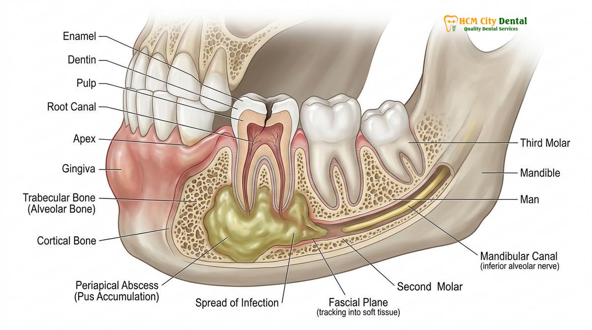
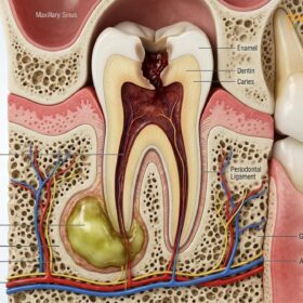
The journey of a severe dental infection typically begins within the hard tissues of the tooth. Dental caries (cavities) that are left untreated eventually penetrate the protective enamel and dentin, reaching the innermost layer known as the dental pulp. The pulp contains the tooth’s nerve and blood supply. Once bacteria infiltrate the pulp, they cause irreversible pulpitis, leading to pulpal necrosis (death of the nerve tissue). Because the tooth is a rigid structure, the accumulating bacteria and their toxic byproducts have only one route of escape: down through the root canal system and out the apical foramen (the tip of the root) into the surrounding alveolar bone.
Once the infection enters the jawbone, it forms a periapical abscess. If the immune system cannot contain this localized pocket of pus, the infection will follow the path of least resistance. It will erode through the cortical plate of the jawbone and spill into the adjacent soft tissues. Depending on which tooth is infected and the specific anatomical attachments of the facial muscles, the infection will dictate where the swelling appears. For example, an infection in an upper canine tooth may spread to the infraorbital space, causing swelling right below the eye. Conversely, an infection in a lower molar often spreads to the submandibular or sublingual spaces, causing swelling under the jaw and pushing the tongue upward.
“The fascial planes of the head and neck act as highways for bacterial dissemination. An odontogenic infection can travel from the mandible to the mediastinum (the chest cavity) in a matter of days if source control is not rapidly achieved.”
This rapid dissemination is what makes a facial swelling dental infection so incredibly dangerous. The head and neck region contains vital structures, most notably the airway. If an infection spreads bilaterally into the submandibular, sublingual, and submental spaces—a condition known clinically as Ludwig’s angina—the massive swelling can elevate the tongue and physically crush the trachea, leading to asphyxiation. Furthermore, the proximity of the upper teeth to the maxillary sinus and the cavernous sinus (a major venous structure at the base of the brain) means that upper jaw infections carry the risk of cavernous sinus thrombosis, a rare but highly fatal complication where a blood clot forms in the brain in response to the infection [2].
Understanding Dental Cellulitis & Abscesses
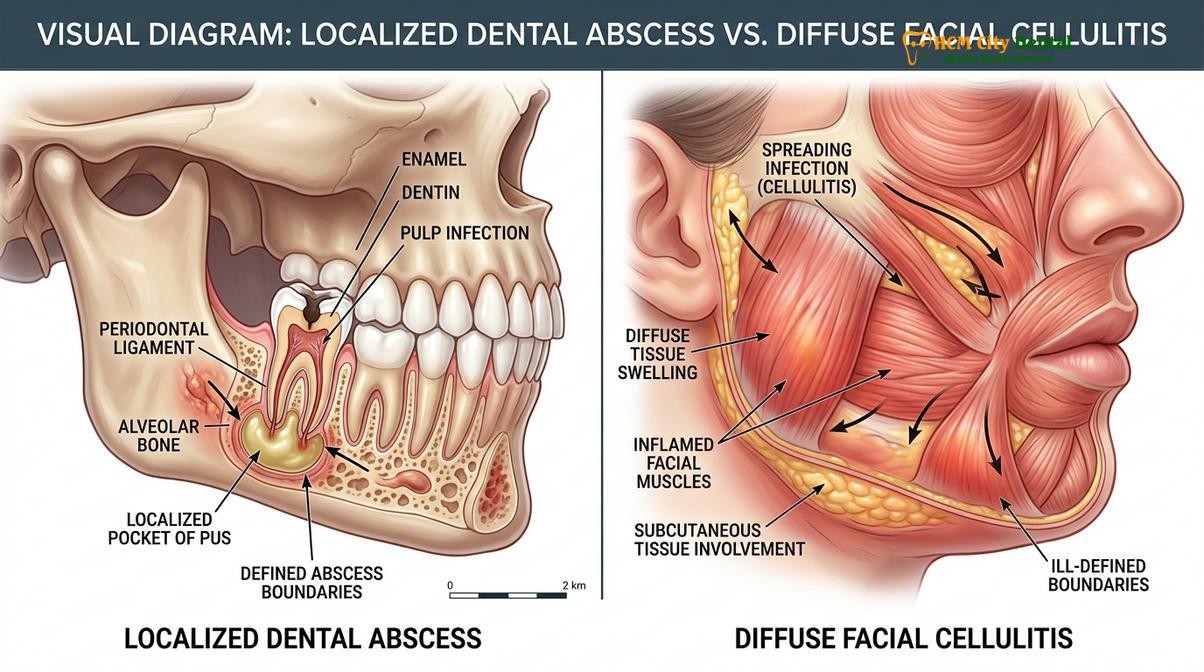
A dental abscess is a localized pocket of pus, whereas dental cellulitis is a diffuse, spreading bacterial infection of the skin and subcutaneous tissues.
When patients present with a swollen face, clinicians must differentiate between two primary stages of infection: the localized abscess and the spreading cellulitis. While both are serious, their clinical presentations, underlying pathophysiology, and immediate treatment requirements differ significantly. Understanding this distinction is crucial for determining the appropriate trajectory of emergency care.
A dental abscess is a localized collection of pus. Pus is a thick, yellowish-white fluid composed of dead white blood cells (neutrophils), tissue debris, and living and dead bacteria. The body forms an abscess as a defense mechanism; it attempts to wall off the infection using a barrier of granulation tissue to prevent the bacteria from spreading systemically. Abscesses are typically fluctuant, meaning they feel like a fluid-filled balloon when palpated by a dentist. They are highly localized, intensely painful due to the extreme pressure building up within the confined space, and often present with a distinct “pointing” head where the pus is trying to rupture through the mucosal surface [3].
In contrast, a cellulitis tooth infection represents a failure of the body’s attempt to wall off the bacteria. Cellulitis is an acute, diffuse, spreading infection of the deep dermis and subcutaneous tissues. Unlike an abscess, cellulitis is not localized to a single pocket. Instead, the bacteria produce enzymes, such as hyaluronidase and fibrinolysin, which break down the connective tissue barriers, allowing the infection to spread rapidly through the tissue planes. Clinically, an abscess face swelling that has progressed to cellulitis will feel firm (indurated) and board-like to the touch, rather than fluid-filled. The overlying skin is typically erythematous (red), warm, and highly tender. Cellulitis is a clear indicator of a highly aggressive, invasive infection that requires immediate systemic intervention.
| Clinical Feature | Dental Abscess | Dental Cellulitis |
|---|---|---|
| Nature of Infection | Localized collection of pus | Diffuse, spreading tissue infection |
| Palpation (Feel) | Fluctuant (fluid-filled, soft) | Indurated (firm, board-like, hard) |
| Boundaries | Well-defined, distinct borders | Poorly defined, spreading borders |
| Pain Characteristics | Intense, localized, throbbing pressure | Widespread tenderness, diffuse pain |
| Primary Treatment | Incision and drainage, source control | Aggressive antibiotics, source control |
The microbiology of these infections is complex and typically polymicrobial, meaning multiple types of bacteria are involved simultaneously. The initial stages of a dental infection are often dominated by aerobic (oxygen-dependent) bacteria, primarily Streptococcus species. However, as the infection progresses deep into the tissues and oxygen levels drop, the environment becomes highly favorable for strictly anaerobic bacteria (bacteria that thrive without oxygen). Species such as Prevotella, Porphyromonas, and Fusobacterium begin to dominate the flora. These anaerobes are highly virulent and are primarily responsible for the tissue destruction, foul odor, and rapid spread characteristic of severe facial cellulitis [4]. This shift in bacterial populations dictates the choice of antibiotics used in emergency settings, necessitating broad-spectrum coverage that specifically targets anaerobic organisms.
Warning Signs: When to Go to the Emergency Room
Seek immediate hospital emergency care if facial swelling is accompanied by difficulty breathing, swallowing, high fever, or swelling that extends to the eye or neck.
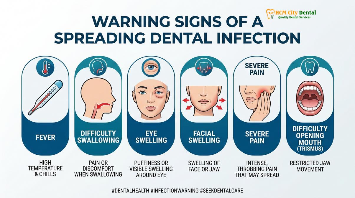
While a localized toothache can often wait for a scheduled dental appointment, a swollen face tooth infection emergency requires immediate triage to determine if the patient needs a dental clinic or a hospital emergency department. Recognizing the red flag symptoms of a spreading infection is critical for patient survival. When an infection breaches the local confines of the jaw, it can rapidly compromise vital systemic functions.
The most critical warning sign is any compromise to the airway. If a patient experiences dyspnea (difficulty breathing), a sensation that their throat is closing, or stridor (a high-pitched wheezing sound when breathing), this is an absolute, life-threatening emergency. This indicates that the swelling has reached the parapharyngeal or retropharyngeal spaces, physically compressing the trachea. In such cases, securing the airway via intubation or tracheostomy takes precedence over all other treatments.
Another severe dental infection spreading sign is dysphagia (difficulty swallowing) or odynophagia (painful swallowing). If a patient cannot swallow their own saliva and begins drooling, it suggests significant involvement of the sublingual and submandibular spaces. The swelling pushes the base of the tongue upward and backward, obstructing the oropharynx. This is a classic presentation of impending Ludwig’s angina and requires immediate hospital admission.
Trismus, commonly known as lockjaw, is a profound inability to open the mouth. While mild stiffness can occur with any jaw pain, severe trismus (opening less than 20 millimeters) indicates that the infection has invaded the masticator space, inflaming the muscles of mastication (chewing). Severe trismus makes it physically impossible for a dentist to access the infected tooth to perform an extraction or root canal, often necessitating treatment under general anesthesia in a hospital setting.
URGENT CLINICAL WARNING: Do not attempt to “wait out” a facial swelling if you experience any of the following systemic symptoms: a high fever (above 101°F / 38.3°C), rapid heart rate (tachycardia), confusion, lethargy, or swelling that rapidly approaches the eye or tracks down the neck. These are signs of systemic inflammatory response syndrome (SIRS) and potential sepsis, requiring immediate intravenous medical intervention.
Swelling that extends upward toward the eye is particularly dangerous. The venous drainage of the upper face lacks valves, meaning blood can flow in both directions. An infection in the upper jaw can travel through the facial vein directly into the cavernous sinus at the base of the brain. Symptoms such as periorbital edema (swelling around the eye), ptosis (drooping eyelid), or changes in vision are critical neurological emergencies. Similarly, swelling that tracks downward into the neck indicates the infection is moving toward the mediastinum (the chest cavity containing the heart and lungs), which carries a very high mortality rate if not intercepted immediately.
Emergency Medical Treatments for Facial Swelling
Treatment involves immediate surgical drainage of the infection source, aggressive antibiotic therapy, and airway management to stabilize the patient and eliminate the bacteria.
When a patient presents with a severe facial swelling dental infection, the clinical workflow must be swift, decisive, and methodical. The primary goal of emergency treatment is twofold: stabilize the patient’s systemic health (airway, breathing, circulation) and achieve immediate source control to halt the progression of the bacteria.
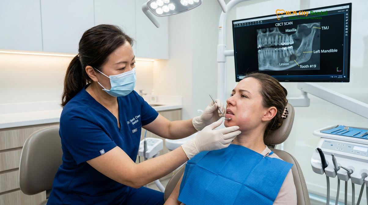
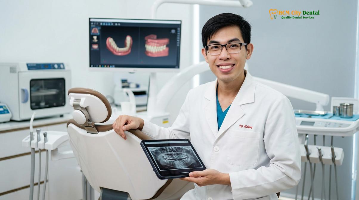
The first step in any severe presentation is a thorough clinical and radiographic assessment. Advanced imaging, such as a Cone Beam Computed Tomography (CBCT) scan or a medical CT scan of the neck with contrast, is often utilized to map the exact anatomical spaces involved and locate the loculations (pockets) of pus. Once the extent of the infection is understood, the clinical team formulates a surgical plan. Dr. Nguyen Van Cuong, a leading expert in complex oral surgical cases, emphasizes that antibiotics alone are never a substitute for physical intervention. The source of the infection—the necrotic tooth—must be addressed.
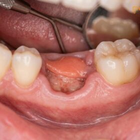
Source control is typically achieved through one of two primary dental procedures: endodontic therapy (root canal) or dental extraction. If the tooth is structurally sound and strategically valuable, an emergency root canal can be initiated. The dentist drills an access hole into the crown of the tooth, entering the pulp chamber. This allows the trapped pus and gas to vent out through the tooth, immediately relieving the internal pressure. The infected nerve tissue is removed, and the canals are thoroughly disinfected with sodium hypochlorite. However, if the tooth is severely decayed, fractured, or lacks sufficient bone support, extraction is the most definitive and rapid method of source control. Removing the tooth completely eliminates the bacterial reservoir and provides a large pathway for the underlying abscess to drain.
In cases where the swelling is extensive and fluctuant, a procedure known as Incision and Drainage (I&D) is performed alongside the tooth treatment. The clinician administers local anesthesia (which can be challenging, as the acidic environment of an infection neutralizes anesthetic agents) and makes a surgical incision directly over the area of maximum swelling. A hemostat (a blunt surgical instrument) is inserted into the incision and opened to break up the loculations of pus, allowing the fluid to evacuate. A small rubber drain may be sutured into the incision site to keep the pathway open for several days, ensuring continuous drainage as the body clears the remaining infection [5].
Clinical Case Review: A 45-year-old patient presented to HCMC Dental Clinic in Ho Chi Minh City with severe, right-sided facial swelling that had developed over 48 hours. The patient exhibited moderate trismus and a low-grade fever. Clinical examination by Dr. Nguyen Van Cuong revealed a necrotic lower right first molar with a massive submandibular abscess. Due to the rapid progression, immediate intervention was required. The patient was placed on broad-spectrum antibiotics, and Dr. Cuong performed an emergency extraction of the offending molar followed by an intraoral incision and drainage. Over 15cc of purulent exudate was evacuated. Within 24 hours, the patient’s swelling had reduced by 60%, and the systemic fever had resolved, highlighting the critical importance of immediate surgical source control.
Pharmacotherapy is the crucial adjunct to surgical treatment. While antibiotics cannot cure an abscess on their own, they are vital for managing the cellulitis component and preventing systemic bacteremia. Empiric antibiotic therapy usually begins with broad-spectrum penicillins, such as Amoxicillin combined with Clavulanate (Augmentin), to overcome bacterial resistance. If anaerobic involvement is severe, Metronidazole may be added to the regimen. For patients with penicillin allergies, Clindamycin or Azithromycin are common alternatives. In hospital settings, intravenous (IV) administration is utilized to achieve rapid, high serum concentrations of the drug. For comprehensive care pathways regarding these urgent scenarios, patients are strongly advised to seek Emergency Dental Care immediately.
What to Do While Waiting for Medical Help
While awaiting professional care, keep your head elevated, apply cold compresses to the outside of the face, and take over-the-counter pain relievers to manage symptoms.
If you are experiencing a swollen face due to a suspected tooth infection, the time between recognizing the symptom and sitting in a dental chair or ER bed can be highly distressing. While you cannot cure the infection at home, there are specific, medically sound steps you can take to manage the pain, slow the progression of swelling, and avoid exacerbating the condition.
First and foremost, keep your head elevated at all times, even when sleeping. Lying flat increases the hydrostatic blood pressure in the head and neck region, which will force more fluid into the inflamed tissues, significantly worsening the swelling and the throbbing pain. Prop yourself up with several thick pillows or sleep in a recliner chair to utilize gravity to your advantage.
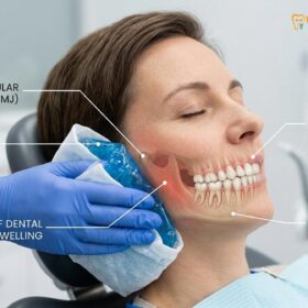
Temperature therapy must be applied correctly. You should only apply cold compresses or ice packs to the outside of the affected cheek. Apply the cold pack for 15 to 20 minutes at a time, followed by a 20-minute break. The cold induces localized vasoconstriction (narrowing of the blood vessels), which helps reduce blood flow to the area, thereby limiting the expansion of the swelling and providing a numbing effect that dulls the pain. Conversely, you must absolutely avoid applying heat to the outside of the face. Heat causes vasodilation (widening of blood vessels), which increases blood flow and can actually draw the infection outward toward the skin surface, potentially causing the abscess to rupture through the cheek—a complication that leaves severe facial scarring.
For pain management, over-the-counter analgesics are your first line of defense. Non-steroidal anti-inflammatory drugs (NSAIDs) like Ibuprofen are highly effective for dental pain because they inhibit the cyclooxygenase (COX) enzymes, thereby reducing the synthesis of prostaglandins, which are the primary chemical mediators of inflammation and pain. Acetaminophen (Paracetamol) can be taken concurrently with Ibuprofen, as they process through different pathways (liver vs. kidneys) and have a synergistic pain-relieving effect. However, always adhere strictly to the maximum daily dosage guidelines on the packaging, and avoid NSAIDs if you have a history of stomach ulcers or bleeding disorders.
Crucially, never attempt to pop, lance, or squeeze the swelling yourself. The fascial spaces are delicate, and applying external pressure can easily rupture the abscess capsule inward, forcing the highly toxic bacteria deeper into the neck or directly into the bloodstream, triggering a massive systemic crisis.
Preventing Tooth Infections from Spreading
Preventing the spread of dental infections requires prompt treatment of early tooth decay, routine dental examinations, and maintaining optimal daily oral hygiene.
The most effective treatment for a severe facial swelling tooth infection is preventing it from occurring in the first place. Dental infections do not materialize overnight; they are the end-stage result of a prolonged, progressive disease process that was left unchecked. Understanding the continuum of dental disease empowers patients to intervene before a minor issue becomes a medical emergency.
The foundation of prevention lies in meticulous daily oral hygiene. Brushing twice daily with a fluoridated toothpaste and flossing once a day disrupts the biofilm (dental plaque) that harbors the bacteria responsible for tooth decay and gum disease. Fluoride plays a critical role by remineralizing weakened enamel, making it more resistant to the acidic byproducts of bacterial metabolism. However, home care alone is insufficient to remove hardened calculus (tartar) or detect microscopic early decay.
“A dental abscess is almost entirely preventable. It represents the final stage of a disease process that typically takes months or years to develop. Early detection through routine radiography is the ultimate safeguard against severe odontogenic infections.”
Routine dental examinations and professional prophylaxis (cleanings) every six months are non-negotiable for optimal oral health. During these visits, clinicians utilize digital X-rays to visualize the interproximal spaces (between the teeth) and the periapical regions (around the roots) that are invisible to the naked eye. Detecting a cavity when it is confined to the enamel or dentin allows for a simple, minimally invasive filling. If a patient ignores the early warning signs—such as sensitivity to cold or sweet foods—the decay will inevitably breach the pulp chamber, necessitating a complex root canal or extraction to prevent an abscess.
At HCMC Dental Clinic, the clinical philosophy heavily emphasizes proactive, preventive care. Patients are educated on the importance of addressing minor dental trauma, such as a cracked or chipped tooth, immediately. Even a microscopic fracture can provide a direct pathway for oral bacteria to invade the sterile pulp chamber. Furthermore, patients with compromised immune systems, such as those with uncontrolled diabetes, must be particularly vigilant, as their bodies are less capable of walling off early infections, making them highly susceptible to rapid, spreading facial cellulitis. By maintaining regular dental visits and addressing issues promptly, patients can ensure that a simple toothache never escalates into a life-threatening emergency.
Frequently Asked Questions
Is facial swelling from a tooth infection dangerous?
Yes, facial swelling from a tooth infection is highly dangerous and potentially life-threatening. It indicates that bacteria have breached the tooth and are spreading into the deep fascial spaces of the head and neck, which can lead to airway obstruction or systemic sepsis. Immediate clinical evaluation is mandatory to prevent these severe complications.
How long can you wait with a swollen face from a tooth?
You should not wait at all; a swollen face from a tooth requires immediate same-day emergency dental or medical care. Odontogenic infections can progress from a localized swelling to a life-threatening airway compromise in a matter of hours, especially in individuals with compromised immune systems. Delaying treatment exponentially increases the risk of requiring hospitalization and invasive surgery.
Will antibiotics alone cure a swollen face from a tooth infection?
No, antibiotics alone cannot cure a swollen face caused by a dental infection. While antibiotics help control the systemic spread of bacteria, they cannot penetrate the source of the infection (the necrotic tooth pulp or abscess cavity). Physical source control, such as root canal therapy, extraction, or surgical drainage, is absolutely required to eliminate the bacterial reservoir.
Can a tooth infection cause sepsis?
Yes, an untreated tooth infection can lead to sepsis. When bacteria from a dental abscess enter the bloodstream (bacteremia), the body may mount an extreme, dysregulated immune response. This systemic inflammatory response can lead to tissue damage, organ failure, and potentially fatal septic shock if not treated aggressively with intravenous antibiotics and source control.
What happens in the ER for a swollen face?
In the emergency room, medical professionals will first secure your airway and assess your vital signs. They may administer intravenous (IV) antibiotics and pain relief, perform a CT scan to map the infection, and potentially drain the abscess. However, you will still need a dentist to treat the underlying infected tooth, as ER physicians generally do not perform root canals or complex extractions.
References
- Journal of Oral and Maxillofacial Surgery. Management of Severe Odontogenic Infections. (2021).
- American Dental Association (ADA). Clinical Guidelines on Emergency Dental Care. (2020).
- International Journal of Dentistry. Pathophysiology of Dental Abscesses and Cellulitis. (2019).
- Journal of Endodontics. Microbiology of Endodontic Infections and Fascial Space Spread. (2022).
- British Journal of Oral and Maxillofacial Surgery. Ludwig’s Angina and Airway Management. (2018).