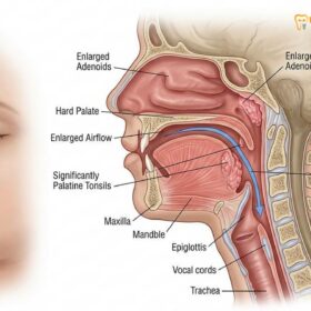
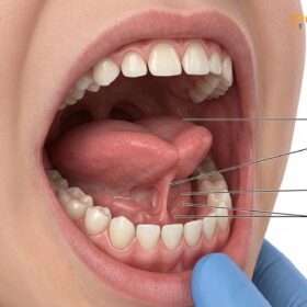
Pediatric sleep apnea is a serious sleep-related breathing disorder characterized by partial or complete upper airway obstruction during sleep. It leads to oxygen desaturation and fragmented sleep, profoundly impacting a child’s neurocognitive development, behavior, and cardiovascular health if left untreated. Early multidisciplinary intervention is essential.
Clinical Summary:
Pediatric obstructive sleep apnea (OSA) is a complex condition driven by anatomical and neuromuscular factors, most notably adenotonsillar hypertrophy and craniofacial anomalies. Unlike adult OSA, pediatric cases frequently manifest as behavioral disturbances, hyperactivity, and learning difficulties, often mimicking ADHD. Diagnosis relies on overnight polysomnography (PSG) evaluated by a qualified sleep physician, as dentists do not diagnose sleep apnea. Treatment requires a collaborative approach involving pediatric ENTs for surgical interventions like adenotonsillectomy, and specialized dentists for structural airway management. Orthodontic treatments, such as Rapid Maxillary Expansion (RME), play a crucial role in widening the nasal floor and decreasing airway resistance. For specific cases, custom oral appliances like the Antisnoring Telescopic or Twinblock Snoring devices can be fabricated to manage airway patency, carefully calibrated through intraoral digital scans and TMJ joint checks to ensure optimal pediatric development.
Key Takeaways:
- Childhood snoring is a primary indicator of sleep-disordered breathing and requires immediate clinical evaluation.
- Behavioral issues, including hyperactivity and poor academic performance, are common neurocognitive consequences of pediatric airway obstruction.
- Diagnosis mandates a formal sleep study (PSG) interpreted by a medical sleep specialist; dentists provide collaborative structural therapies.
- Rapid Maxillary Expansion (RME) is an evidence-based orthodontic therapy used to increase nasal cavity volume and improve airflow.
- Dentists collaborate with sleep physicians to provide custom oral appliances, ensuring proper titration and TMJ safety for growing children.
Recognizing the signs of pediatric airway obstruction and mouth breathing
Recognizing pediatric airway obstruction involves identifying nocturnal symptoms like habitual snoring and gasping, alongside daytime signs such as chronic mouth breathing and fatigue. Early detection prevents long-term craniofacial and systemic complications.
The clinical presentation of pediatric sleep apnea differs significantly from that of adults. While adults often present with excessive daytime sleepiness and loud, intermittent snoring, children may exhibit a wider, more subtle spectrum of symptoms that can easily be overlooked or misattributed to other developmental phases. The hallmark of pediatric obstructive sleep apnea (OSA) is increased upper airway resistance during sleep, which leads to a cascade of physiological disruptions. Parents and caregivers are typically the first to notice nocturnal symptoms, which include habitual snoring, paradoxical chest movements, witnessed apneas (pauses in breathing), gasping, and restless sleep. Children with OSA often adopt unusual sleeping positions, such as hyperextending their necks to open the airway or sleeping seated upright.
Beyond nocturnal symptoms, daytime manifestations are equally critical for clinical screening. Chronic pediatric mouth breathing is one of the most prominent daytime indicators of underlying airway compromise. When the nasal passage is obstructed—whether due to allergic rhinitis, septal deviation, or enlarged lymphoid tissues—the child is forced to breathe through the mouth to maintain adequate oxygenation. This continuous mouth breathing bypasses the nasal cavity’s natural filtration, humidification, and warming functions, leading to chronic dry mouth, increased susceptibility to dental caries, and gingival inflammation. More importantly, chronic mouth breathing alters the resting posture of the tongue. Instead of resting against the palate to stimulate normal lateral maxillary growth, the tongue drops to the floor of the mouth. Over time, this lack of palatal stimulation results in a narrow, high-arched palate, a retrognathic (recessed) mandible, and a lengthened facial profile—a condition clinically referred to as “long face syndrome” or adenoid facies [1].

The pathophysiology of pediatric airway obstruction is primarily driven by the disproportionate growth of lymphoid tissues relative to the size of the pediatric airway. Between the ages of 2 and 8, the tonsils and adenoids undergo significant hypertrophy, often peaking in size just as the child’s airway is still relatively narrow. When the child enters deep sleep, particularly Rapid Eye Movement (REM) sleep, the neuromuscular tone of the pharyngeal dilator muscles decreases. This relaxation, combined with the physical bulk of the enlarged tonsils adenoids, leads to partial or complete collapse of the airway. The resulting obstruction causes intermittent oxygen desaturation and hypercapnia (elevated carbon dioxide levels), triggering the brain to initiate micro-arousals to restore airway patency. These repeated arousals severely fragment the child’s sleep architecture, depriving them of the restorative deep sleep necessary for healthy somatic growth and cognitive development.
“The American Academy of Pediatrics emphasizes that habitual snoring in children is a primary marker for sleep-disordered breathing and warrants comprehensive screening for obstructive sleep apnea to prevent irreversible neurocognitive and cardiovascular sequelae.”
Clinical evaluation of these signs requires a meticulous approach. Dental professionals play a vital role in the early identification of craniofacial risk factors during routine examinations. By assessing the tonsillar grade (using the Brodsky scale), evaluating the transverse width of the maxilla, and noting signs of bruxism (teeth grinding) which is often a subconscious mechanism to bring the jaw forward and open the airway, dentists can identify at-risk children. However, it is imperative to understand that the definitive diagnosis of pediatric sleep apnea relies on objective data obtained through a sleep study, specifically measuring the Apnea-Hypopnea Index (AHI). In pediatric medicine, an AHI of greater than 1 event per hour is considered abnormal and warrants clinical intervention, highlighting the lower threshold for diagnosing respiratory compromise in children compared to adults.
Why child snoring is never normal: Impact on behavioral development and ADHD
Snoring in children indicates increased airway resistance that fragments sleep architecture, leading to severe neurocognitive deficits. These sleep disruptions frequently manifest as hyperactivity, impulsivity, and inattention, closely mimicking ADHD.
A pervasive misconception among parents and even some healthcare providers is that light snoring in children is a benign, temporary condition. In reality, pediatric snoring is an acoustic manifestation of turbulent airflow through a narrowed upper airway. This increased airway resistance forces the child’s respiratory system to work harder, leading to increased intrathoracic pressure and physiological stress. When snoring progresses to obstructive sleep apnea, the consequences extend far beyond the respiratory system, profoundly impacting the child’s developing brain. The dual insults of sleep fragmentation and intermittent hypoxia (drops in blood oxygen levels) disrupt the delicate processes of neurogenesis, synaptic pruning, and memory consolidation that occur during healthy sleep.
The prefrontal cortex, the region of the brain responsible for executive functions such as impulse control, attention regulation, and complex problem-solving, is particularly vulnerable to the oxidative stress caused by intermittent hypoxia. When a child experiences repeated oxygen desaturation events throughout the night, the prefrontal cortex cannot function optimally during the day. Consequently, children with untreated sleep apnea frequently exhibit significant behavioral and cognitive morbidities. Unlike adults who typically present with lethargy and daytime somnolence, children often exhibit a paradoxical reaction to sleep deprivation: hyperactivity. This hyperactive behavior is a compensatory mechanism to stay awake and alert despite profound underlying fatigue [2].
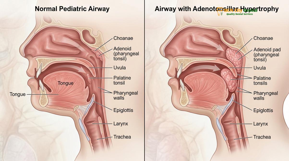
This clinical presentation leads to a critical diagnostic overlap. The symptoms of pediatric sleep apnea—inattention, impulsivity, emotional lability, poor academic performance, and hyperactivity—are virtually indistinguishable from the diagnostic criteria for Attention-Deficit/Hyperactivity Disorder (ADHD). Numerous clinical studies have demonstrated a high prevalence of sleep-disordered breathing among children diagnosed with ADHD. Unfortunately, if the underlying airway obstruction is not identified, these children may be prescribed stimulant medications. While stimulants may temporarily mask the daytime symptoms by artificially increasing alertness, they do nothing to resolve the nocturnal airway collapse and may even exacerbate sleep onset insomnia, further compounding the child’s sleep debt.
Furthermore, the systemic impact of untreated pediatric sleep apnea extends to cardiovascular and metabolic health. The repeated sympathetic nervous system surges triggered by apneic events and micro-arousals lead to elevated blood pressure and altered heart rate variability. Over time, this chronic sympathetic overactivity can predispose the child to early-onset hypertension and endothelial dysfunction. Metabolically, sleep fragmentation disrupts the normal secretion of growth hormone (which is primarily released during slow-wave sleep) and alters the balance of appetite-regulating hormones like leptin and ghrelin, increasing the risk of childhood obesity. Therefore, addressing child snoring is not merely a matter of improving sleep quality; it is a critical intervention to safeguard the child’s holistic developmental trajectory.
Rapid Maxillary Expansion (RME): Orthodontic widening to open nasal airways
Rapid Maxillary Expansion is a highly effective orthodontic intervention that widens the upper jaw, thereby expanding the nasal floor and significantly reducing upper airway resistance in growing children.
When anatomical narrowing of the maxilla contributes to pediatric sleep apnea, structural intervention is required to permanently alter the airway geometry. Rapid Maxillary Expansion (RME) is an evidence-based orthodontic procedure designed to correct transverse maxillary deficiency. The maxilla (upper jaw) forms the floor of the nasal cavity and the roof of the mouth. In children with chronic mouth breathing or genetic predispositions, the maxilla often develops in a narrow, V-shape rather than a broad, U-shape. This transverse discrepancy not only leads to dental crowding and crossbites but also physically constricts the nasal airway, increasing nasal resistance and forcing the child to breathe through their mouth.
The biomechanical principle of RME relies on the fact that in growing children, the midpalatal suture—the fibrous joint connecting the two halves of the maxilla—has not yet fused. By applying controlled, heavy orthopedic forces to the teeth and the palatal vault using a custom-fabricated expansion appliance, orthodontists can separate the two halves of the maxilla. As the suture opens, new bone is generated in the gap through a process called distraction osteogenesis. Because the maxilla forms the floor of the nasal cavity, widening the maxilla simultaneously widens the nasal floor, significantly increasing the total volume of the nasal airway and decreasing airflow resistance [3].
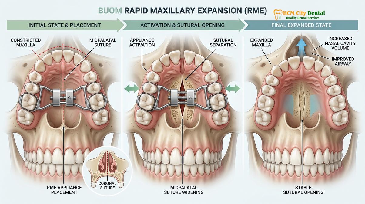
From a clinical perspective, Dr. Nguyen Van Cuong emphasizes the importance of precise airway scanning and intraoral digital scans to evaluate the transverse discrepancy before initiating rapid maxillary expansion. At HCMC Dental Clinic, the diagnostic workflow involves comprehensive 3D imaging to assess the exact dimensions of the nasal cavity, the thickness of the palatal bone, and the stage of skeletal maturation. This data allows the dental team to customize the expansion protocol, ensuring that the forces applied maximize skeletal expansion while minimizing unwanted dental tipping. The RME appliance is typically activated daily by the parents using a small key, gradually widening the palate over a period of two to four weeks. Following the active expansion phase, the appliance is left in place for several months to act as a rigid retainer, allowing the newly formed bone in the midpalatal suture to mineralize and stabilize.
| Intervention Type | Primary Indication | Mechanism of Action | Clinical Considerations |
|---|---|---|---|
| Rapid Maxillary Expansion (RME) | Transverse maxillary deficiency, narrow nasal floor | Orthopedic separation of the midpalatal suture to increase nasal volume. | Most effective before the midpalatal suture fuses (typically ages 7-12). |
| Adenotonsillectomy | Severe adenotonsillar hypertrophy | Surgical removal of obstructing lymphoid tissues in the pharynx. | First-line medical treatment for severe pediatric OSA; requires ENT evaluation. |
| Custom Oral Appliances | Mild-to-moderate OSA, residual snoring post-surgery | Mandibular advancement to prevent posterior tongue collapse. | Requires careful titration and TMJ joint checks to ensure healthy growth. |
The clinical outcomes of RME in the context of pediatric sleep apnea are profound. By restoring normal nasal breathing, RME eliminates the primary trigger for mouth breathing. This allows the tongue to return to its proper resting position against the palate, which naturally supports the airway and prevents the tongue from collapsing backward during sleep. Furthermore, the reduction in nasal resistance decreases the negative intrathoracic pressure required to inhale, thereby reducing the likelihood of pharyngeal collapse. Clinical studies have consistently shown that RME can significantly reduce the Apnea-Hypopnea Index (AHI) and improve oxygen saturation in children with maxillary constriction and OSA. However, RME is most effective when utilized as part of a comprehensive treatment plan that may also include surgical or myofunctional therapies to address all levels of airway obstruction.
Collaborating with pediatric ENTs for tonsillectomy and adenoidectomy
For children with significant adenotonsillar hypertrophy, surgical removal of the tonsils and adenoids remains the first-line medical treatment to resolve upper airway obstruction and normalize breathing.
While orthodontic interventions like RME address the structural dimensions of the bony airway, soft tissue obstructions require a different clinical approach. In the pediatric population, the most common etiology of obstructive sleep apnea is the enlargement of the palatine tonsils and the pharyngeal tonsils (adenoids). When these lymphoid tissues become hypertrophic—often due to recurrent infections, allergies, or genetic factors—they physically occlude the oropharynx and nasopharynx. In such cases, the American Academy of Pediatrics and the American Academy of Otolaryngology-Head and Neck Surgery recommend adenotonsillectomy (the surgical removal of the tonsils and adenoids) as the first-line treatment for pediatric OSA.
The management of pediatric sleep apnea is inherently multidisciplinary. Dentists, orthodontists, and sleep physicians must collaborate closely with pediatric Ear, Nose, and Throat (ENT) specialists to ensure comprehensive care. When a child presents to a dental clinic with signs of severe airway obstruction, such as a Brodsky Grade 3 or 4 tonsillar hypertrophy, the immediate clinical protocol involves a referral to an ENT for surgical evaluation. The ENT specialist will perform a thorough endoscopic examination of the upper airway to assess the degree of obstruction and determine the child’s candidacy for surgery. Following a formal diagnosis of OSA via polysomnography, the adenotonsillectomy is performed under general anesthesia to mechanically clear the airway [4].
“A multidisciplinary approach, integrating otolaryngology, dental sleep medicine, and myofunctional therapy, yields the highest success rates in achieving complete resolution of pediatric sleep-disordered breathing and preventing relapse.”
However, the clinical journey does not end in the operating room. While adenotonsillectomy is highly effective, resolving OSA in approximately 70% to 80% of healthy children, a significant subset of patients experiences residual sleep-disordered breathing post-surgery. This residual obstruction often occurs because the child has developed ingrained neuromuscular habits, such as chronic mouth breathing and low tongue posture, which persist even after the anatomical blockage is removed. This is where the collaboration between the ENT and the dental professional becomes critical. Post-surgical dental evaluations are necessary to assess whether the child requires subsequent orthodontic expansion or myofunctional therapy. Myofunctional therapy involves specific exercises designed to retrain the orofacial muscles, promote nasal breathing, and establish proper tongue posture, thereby ensuring the long-term stability of the surgical and orthodontic outcomes.
Custom Oral Appliances for Sleep Apnea & Snoring Management
When surgery is contraindicated or residual mild-to-moderate OSA persists, custom oral appliances fabricated by specialized dentists offer a non-invasive solution to maintain airway patency during sleep.
In scenarios where a child has residual sleep apnea following an adenotonsillectomy, or when surgery is contraindicated, custom oral appliances serve as a vital therapeutic option. These devices, known as Mandibular Advancement Devices (MADs), work by gently repositioning the lower jaw (mandible) forward during sleep. This forward movement pulls the base of the tongue away from the posterior pharyngeal wall, tightening the soft tissues and muscles of the upper airway, thereby preventing collapse and maintaining continuous airflow. It is crucial to reiterate that dentists do not diagnose sleep apnea; the fabrication of these devices is always done in collaboration with a medical sleep physician who has formally diagnosed the condition and prescribed oral appliance therapy.
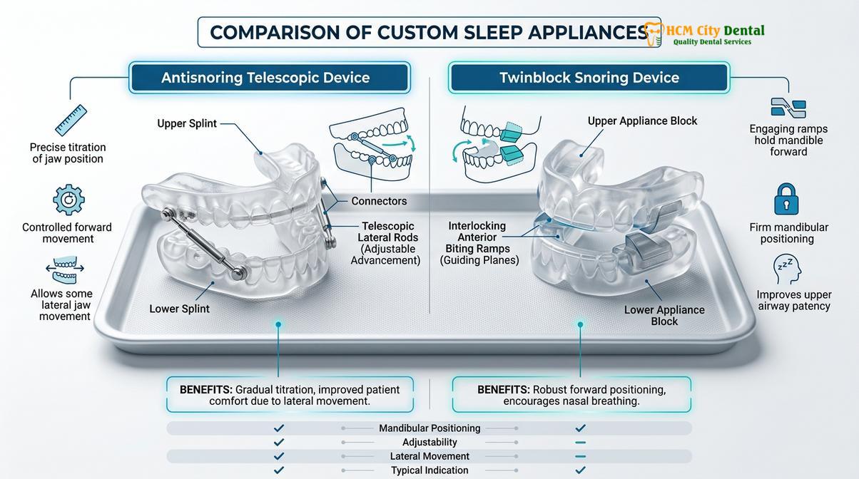
At HCMC Dental Clinic, the clinical protocol for fabricating custom oral appliances for sleep-disordered breathing involves utilizing state-of-the-art intraoral digital scans to capture the exact topography of the patient’s dentition. This digital precision ensures a perfect fit, which is essential for patient compliance and comfort. The clinic offers specific, highly engineered devices tailored to the patient’s anatomical needs:
- Antisnoring Telescopic Device: This appliance utilizes a Herbst-style mechanism featuring lateral telescopic rods. This advanced design allows for both side-to-side and vertical jaw movement while maintaining the forward posture of the mandible. By permitting this range of motion, the device significantly minimizes temporomandibular joint (TMJ) stiffness and discomfort, making it highly suitable for patients who may experience claustrophobia with rigid devices. Currently, this premium device is priced at 12,000,000 VND (approximately $480 USD with a special WhatsApp discount).
- Twinblock Snoring Device: This is a two-piece appliance consisting of upper and lower plates with interlocking 70-degree bite blocks. When the patient closes their mouth, the angled blocks guide the lower jaw into a predetermined forward position. The Twinblock is highly effective for growing patients as it not only manages the airway but can also assist in correcting skeletal Class II malocclusions (overbites). This device is priced at 11,000,000 VND (approximately $440 USD with a special WhatsApp discount).
The success of oral appliance therapy in pediatric and adolescent patients hinges on meticulous clinical oversight. Dr. Cuong meticulously oversees the titration calibration—the gradual advancement of the mandible to the optimal therapeutic position that resolves apneas without causing muscular strain. Furthermore, because children are actively growing, Dr. Cuong conducts thorough TMJ joint checks at regular intervals. These checks are critical to ensure that the appliance supports healthy craniofacial growth without causing condylar displacement, joint dysfunction, or unwanted shifts in the dental occlusion [5]. By combining advanced digital fabrication with rigorous clinical monitoring, the dental team provides a safe, effective, and highly personalized approach to airway management.
When to See a Doctor for Pediatric Sleep Apnea
Identifying the right time to seek medical intervention is crucial for preventing the long-term complications of pediatric sleep apnea. Parents should immediately consult a healthcare professional if they observe red flag symptoms such as witnessed pauses in breathing during sleep, gasping or choking noises, severe nocturnal sweating, or episodes of cyanosis (bluish discoloration of the lips or face). Additionally, if a child exhibits chronic daytime fatigue, unexplained behavioral issues, hyperactivity, or a decline in academic performance alongside habitual snoring, a comprehensive sleep evaluation is strongly indicated.
Important Clinical Considerations and Medical Disclaimer: It is imperative to understand that dentists DO NOT diagnose sleep apnea. The definitive diagnosis of obstructive sleep apnea requires an objective sleep study, such as overnight Polysomnography (PSG) or a Home Sleep Apnea Test (HSAT), which must be evaluated and interpreted by a board-certified sleep physician. The role of the dental professional is strictly collaborative. Dentists work in tandem with sleep physicians and ENTs to fabricate custom oral appliances (MADs) for patients with mild-to-moderate OSA, those who are CPAP-intolerant, or individuals suffering from primary snoring. Any structural intervention, including rapid maxillary expansion or appliance therapy, requires a personalized clinical examination to determine candidacy, rule out contraindications (such as severe TMJ disorders or insufficient dentition), and ensure the treatment aligns with the child’s overall medical and developmental needs.
Frequently Asked Questions

Is mouth breathing in children a sign of sleep apnea?
Yes, chronic pediatric mouth breathing is a strong clinical indicator of upper airway resistance and potential sleep apnea. It suggests that the nasal airway is obstructed, often due to enlarged tonsils adenoids or a narrow maxilla. Continuous mouth breathing alters facial growth, leading to a high-arched palate and retrognathic mandible, which further exacerbates airway collapsibility during sleep and requires comprehensive medical and dental evaluation.
Can pediatric braces help expand a child’s airway?
Yes, specific orthodontic treatments like rapid maxillary expansion can significantly increase the volume of the nasal cavity and airway. By widening the upper jaw before the midpalatal suture fuses, orthodontists can decrease nasal airway resistance. This structural change improves nasal breathing, facilitates proper tongue posture, and reduces the severity of sleep-disordered breathing in growing children.
How does Dr. Cuong screen pediatric airways?
Dr. Cuong utilizes comprehensive clinical evaluations, including intraoral digital scans and 3D imaging, to assess craniofacial structures and airway dimensions. During consultations at HCMC Dental Clinic, he examines palate width, tonsillar hypertrophy, and jaw relationships. If structural narrowing is detected, he collaborates with medical sleep specialists for a formal diagnosis and coordinates appropriate orthodontic or appliance therapy.
What is the difference between primary snoring and pediatric sleep apnea?
Primary snoring involves noisy breathing without significant oxygen desaturation or sleep fragmentation, whereas sleep apnea includes actual breathing pauses and physiological stress. While primary snoring is considered less severe, it still indicates airway resistance and can progress to obstructive sleep apnea. Both conditions require clinical evaluation to rule out underlying anatomical issues and prevent long-term neurocognitive impacts.
At what age can a child start treatment for sleep apnea?
Treatment can begin as early as ages 2 to 5, which is the peak age for adenotonsillar hypertrophy and related airway obstruction. Surgical interventions like adenotonsillectomy are common in this age group. Orthodontic interventions, such as rapid maxillary expansion, are typically initiated between ages 7 and 10, taking advantage of the child’s active craniofacial growth to permanently widen the airway.
References
- American Academy of Pediatrics. Clinical Practice Guideline: Diagnosis and Management of Childhood Obstructive Sleep Apnea Syndrome. (2012).
- Journal of Clinical Sleep Medicine. Neurocognitive and Behavioral Morbidity in Children with Sleep Disorders. (2018).
- American Journal of Orthodontics and Dentofacial Orthopedics. Effects of rapid maxillary expansion on the upper airway. (2020).
- Otolaryngology–Head and Neck Surgery. Multidisciplinary management of pediatric sleep-disordered breathing. (2019).
- Journal of Dental Sleep Medicine. Efficacy of custom oral appliances in pediatric and adolescent populations. (2021).
For professional children’s dental care, preventive sealants, or to schedule a check-up, visit our Children & Pediatric Dentistry service page at HCMC Dental Clinic in Ho Chi Minh City.