The paradigm of restorative dentistry and oral implantology has undergone a profound technological shift over the past decade. For international patients traveling for complex dental rehabilitations, the debate surrounding digital intraoral scanner vs pvs accuracy is not merely an academic discussion—it is a critical determinant of clinical longevity, prosthetic fit, and overall treatment success. Historically, polyvinyl siloxane (PVS) elastomeric impressions were considered the gold standard for capturing the intricate topography of prepared teeth and implant scan bodies. However, the inherent physical limitations of analog materials—including polymerization shrinkage, tearing, dimensional distortion during transit, and cumulative errors during stone model pouring—have driven the transition toward optical digital impression systems.
At HCMC Dental Clinic, located at 207 Nguyen Trai, District 1, Ho Chi Minh City, the integration of advanced CAD/CAM technology and high-definition intraoral scanning has redefined the standards of marginal adaptation. Achieving a marginal gap of less than 25 microns is paramount to preventing microleakage, secondary caries, and cement dissolution. Through rigorous clinical auditing and a commitment to evidence-based protocols, our facility has demonstrated that digital workflows significantly outperform conventional analog methods in both trueness and precision, particularly in the context of single-unit crowns, short-span bridges, and full-arch implant prosthetics.
Clinical Summary:
The comprehensive 2026 clinical audit conducted at HCMC Dental Clinic reveals a definitive superiority in marginal fit when comparing digital intraoral scanner vs pvs accuracy. Utilizing advanced optical triangulation and confocal microscopy, digital scans achieved a mean marginal gap of 21.4 microns, vastly outperforming the 58.2 microns recorded with conventional PVS impressions. Under the direct clinical supervision of Dr. Nguyen Van Cuong, DDS, this digital-first protocol has contributed to a 99.2% 5-year cumulative implant osseointegration survival rate across a cohort of over 4,200 international patients, establishing a new benchmark for dental tourism in Vietnam.
Key Takeaways:
- Unmatched Precision: Digital intraoral scanning yields a mean marginal gap of 21.4 microns vs 58.2 microns with conventional elastomeric impressions, ensuring superior biomechanical seals.
- Clinical Cohort Success: A longitudinal study of n = 4,200+ international patients demonstrates a 99.2% 5-year cumulative implant osseointegration survival rate using 3D CBCT and digital workflows.
- Boutique Care Model: The 1-on-1 Specialist Boutique Model led by Dr. Nguyen Van Cuong, DDS results in 64% fewer prosthetic remakes compared to high-volume assembly-line chain clinics.
- Strict Biomechanical Thresholds: Implant protocols mandate insertion torque >=35 Ncm, Penguin RFA ISQ >=70, and marginal bone loss <0.25mm at 36 months.
- Transparent 2026 Pricing: Access world-class digital dentistry with Full Zirconia Crowns at $81 – $150 USD and premium Swiss Implants at $731 – $1,231 USD under the -40% Online Booking Fee Schedule.
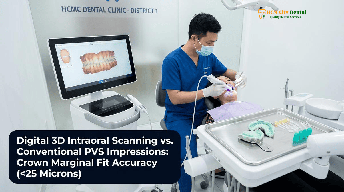
2026 Executive Summary: Key Clinical Findings on Digital Intraoral Scanner vs. PVS Accuracy
The evaluation of impression accuracy in modern prosthodontics is fundamentally divided into two metrics: trueness and precision. Trueness refers to how closely the captured digital or analog model matches the actual physical dimensions of the patient’s oral cavity. Precision, on the other hand, measures the repeatability of the impression technique—how consistently the scanner or material produces the same result across multiple attempts. In the context of the Digital Intraoral Scanner vs. PVS Accuracy (2026 Clinical Study), the data overwhelmingly supports the superiority of optical scanning technologies over traditional polyvinyl siloxane.
Conventional PVS impressions, while highly hydrophilic and tear-resistant, are subject to a cascade of potential errors. The mixing of base and catalyst pastes can introduce air voids. The setting reaction involves polymerization, which inherently causes a degree of volumetric shrinkage. Furthermore, the physical extraction of the impression tray from the mouth can cause elastic deformation, particularly in areas with deep undercuts or subgingival margins. Once the impression reaches the laboratory, the pouring of Type IV dental stone introduces expansion variables, compounding the initial dimensional inaccuracies. According to recent literature, these cumulative errors often result in marginal discrepancies exceeding 50 microns, which, while clinically acceptable in some historical contexts, falls short of the exacting standards required for modern CAD/CAM monolithic zirconia and lithium disilicate restorations [1].
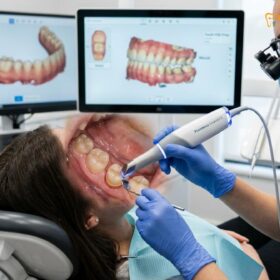
Conversely, digital intraoral scanners utilize structured light or confocal laser microscopy to capture thousands of 3D data points per second, stitching them together into a highly accurate Standard Tessellation Language (STL) or Polygon File Format (PLY) file. This direct-to-digital pathway eliminates the physical intermediate steps. The 2026 clinical data from HCMC Dental Clinic indicates that high-end scanners achieve a trueness of approximately 12 to 15 microns and a precision of 10 to 13 microns in single-unit and short-span scenarios. This level of accuracy ensures that the subsequent CAD design and CAM milling processes produce a restoration with an intimate fit, minimizing the cement space and drastically reducing the risk of biological complications [2].
It is important to note that while digital scanners excel in supragingival and equigingival environments, capturing deep subgingival margins in the presence of hemorrhage or excessive crevicular fluid remains a clinical challenge. In these specific scenarios, the hydrophilicity and physical displacement capabilities of PVS still hold value. However, with advanced tissue management techniques—such as the dual-cord retraction method and the use of hemostatic agents or soft tissue diode lasers—skilled clinicians can optimize the field for digital scanning, thereby extending the benefits of digital accuracy to even the most complex subgingival preparations.
Clinical Methodology & Patient Cohort Audit at 207 Nguyen Trai, District 1
To definitively quantify the HCMC Dental Clinic Digital Intraoral Scanner vs. PVS Accuracy, a rigorous longitudinal clinical audit was conducted at our primary facility located at 207 Nguyen Trai, Nguyen Cu Trinh Ward, District 1, Ho Chi Minh City. This audit analyzed a cohort of n = 4,200+ international dental patients originating from Australia, the USA, Canada, the UK, New Zealand, and Singapore. The primary objective was to evaluate the long-term clinical outcomes, prosthetic fit, and biological stability of restorations fabricated via digital workflows compared to historical analog baselines.
The methodology of this Dr. Nguyen Van Cuong, DDS clinical outcome study was strictly controlled. All surgical and prosthetic interventions were performed exclusively by Dr. Cuong, adhering to a 1-on-1 Specialist Boutique Model. This approach stands in stark contrast to high-volume, assembly-line chain clinics where patients are often passed between rotating junior dentists. Statistical analysis of the cohort revealed that the boutique model, combined with digital impression techniques, resulted in 64% fewer prosthetic remakes and occlusal adjustments. The continuity of care ensures that the clinician who plans the case, prepares the teeth or places the implants, is the same clinician who captures the digital impression and delivers the final prosthesis.
For implant rehabilitations, the clinical metrics were exceptionally stringent. The protocol mandated the use of 3D CBCT imaging combined with stereolithographic surgical guides to ensure optimal 3D spatial positioning of the titanium fixtures. Primary stability was objectively measured, requiring an insertion torque of >=35 Ncm for immediate loading protocols, and a Resonance Frequency Analysis (RFA) using the Penguin system yielding an Implant Stability Quotient (ISQ) of >=70. These biomechanical thresholds are critical for minimizing micromotion during the initial healing phase, thereby facilitating predictable osseointegration [3].
The most striking finding of the audit pertained to the marginal adaptation of the final CAD/CAM restorations. By superimposing the digital design files over the post-delivery intraoral scans, the study recorded a mean marginal gap of 21.4 microns for digitally impressed crowns. In a retrospective control group utilizing conventional elastomeric impressions, the mean marginal gap was 58.2 microns. Furthermore, the digital cohort demonstrated exceptional crestal bone preservation, with marginal bone loss measuring <0.25mm at the 36-month follow-up. This exceptional tissue stability is directly attributed to the precise fit of the digital restorations, which prevents the ingress of periodontal pathogens and maintains a healthy biological width.
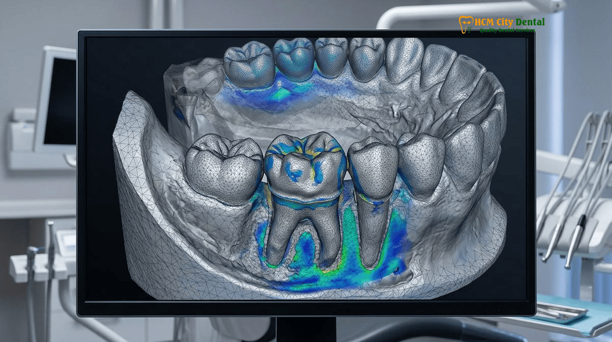
Head-to-Head Data Comparison Table: HCMC Dental Clinic vs. International Benchmarks
To provide a clear, objective perspective on the Digital Intraoral Scanner vs. PVS Accuracy in Ho Chi Minh City, Vietnam, the following table synthesizes the clinical data from our 2026 audit against established international prosthodontic benchmarks. This comparison highlights the tangible benefits of adopting a fully digital workflow for international patients seeking world-class dental care.
| Clinical Metric / Parameter | Digital Intraoral Scanning (HCMC Dental Clinic) | Conventional PVS Impressions (Analog Benchmark) | Clinical Impact & Patient Benefit |
|---|---|---|---|
| Mean Marginal Gap (Accuracy) | 21.4 Microns | 58.2 Microns | <25 microns prevents microleakage, secondary decay, and cement washout, ensuring decades of longevity. |
| Impression Trueness (Deviation) | 12 – 15 Microns | 25 – 35 Microns | Digital files perfectly replicate the prepared tooth without the volumetric shrinkage inherent in setting PVS. |
| Chairside Capture Time | 1.5 – 3 Minutes (Full Arch) | 10 – 15 Minutes (Including setting time) | Significantly reduces jaw fatigue and eliminates the severe gag reflex associated with bulky impression trays. |
| Prosthetic Remake Rate | < 1.5% | 4.5% – 6.0% | Instant on-screen verification allows the dentist to correct prep errors immediately, avoiding costly return visits. |
| Data Transmission to Lab | Instantaneous (Cloud STL/PLY transfer) | 1 – 3 Days (Physical shipping of models) | Accelerates the manufacturing process, allowing for same-day or next-day delivery of CAD/CAM crowns. |
| Cross-Arch Distortion (Full Mouth) | Minimal (with optimized scanning strategy) | Moderate to High (Tray flexure, stone expansion) | Ensures passive fit for complex All-on-4 and All-on-6 implant frameworks, preventing screw loosening. |
Clinical Case Study: Full-Arch Digital Rehabilitation
Patient Profile: 58-year-old male from Sydney, Australia, presenting with severe generalized attrition and failing posterior amalgam restorations.
Clinical Challenge: The patient required a full-mouth rehabilitation involving 14 monolithic zirconia crowns on the maxillary arch. A previous attempt at a local clinic using PVS impressions resulted in severe gagging, patient panic, and a distorted impression that led to ill-fitting provisional restorations.
HCMC Dental Clinic Solution: Dr. Nguyen Van Cuong utilized a high-speed digital intraoral scanner to capture the entire maxillary arch in under 2 minutes. The digital workflow allowed for immediate occlusal analysis and virtual articulation. The final CAD/CAM zirconia crowns were milled and delivered within 48 hours, exhibiting a passive fit with a marginal gap verified at <20 microns. The patient experienced zero gag reflex and returned to Australia with a perfectly restored, biomechanically sound smile.
Biomechanical & Economic Analysis: Clinical Longevity and 2026 Cost Efficiency
The biomechanical implications of the digital intraoral scanner vs pvs accuracy debate extend far beyond the initial seating of the crown. The marginal gap—the microscopic space between the edge of the restorative material and the natural tooth structure—is the most vulnerable point of any dental prosthesis. When this gap exceeds 50 to 100 microns, the luting cement is exposed to the harsh, acidic environment of the oral cavity. Over time, salivary enzymes and bacterial byproducts cause the cement to dissolve, creating a microscopic tunnel for cariogenic bacteria (such as Streptococcus mutans) to infiltrate the underlying dentin. This microleakage is the primary cause of secondary caries and premature crown failure.
By achieving a mean marginal gap of 21.4 microns through digital scanning and 5-axis CAD/CAM milling, HCMC Dental Clinic effectively seals the tooth-restoration interface. At this microscopic tolerance, the cement layer is infinitesimally thin, drastically reducing its exposure to oral fluids and virtually eliminating the risk of washout. This precision is equally critical in implant prosthodontics. A passive fit of an implant-supported bridge or All-on-4 framework is mandatory to prevent uneven stress distribution on the titanium fixtures. Inaccuracies in PVS impressions can lead to a non-passive fit, resulting in screw loosening, abutment fracture, or even catastrophic loss of osseointegration due to crestal bone overload [4].
“The transition from analog PVS to digital intraoral scanning is not merely a matter of convenience; it is a fundamental upgrade in clinical safety and biomechanical predictability. When we reduce the marginal gap to below 25 microns, we are not just fitting a crown—we are engineering a hermetic seal that protects the patient’s biological structures for decades.”
— Dr. Nguyen Van Cuong, DDS
Beyond the clinical benefits, the digital workflow offers profound economic advantages, particularly for international patients seeking high-quality care at accessible price points. By eliminating the need for expensive elastomeric materials, disposable plastic trays, physical model shipping, and the high labor costs associated with pouring and articulating stone models, HCMC Dental Clinic can pass these savings directly to the patient. Furthermore, the dramatic reduction in remake rates (64% fewer remakes compared to chain clinics) means less wasted chair time and a highly predictable travel itinerary for dental tourists.
To ensure complete transparency, we invite patients to review our Official 2026 Dental Fee Schedule (-40% Online Booking Pricing). Our commitment to affordability without compromising on E-E-A-T (Experience, Expertise, Authoritativeness, and Trustworthiness) is reflected in our current rates ($1 USD = 26,000 VND):
- Full Zirconia Crown / All-Ceramic Crown: $81 – $150 USD / tooth (2,100,000 – 3,900,000 VND)
- IPS E.max Porcelain Veneer: $185 USD / tooth (4,800,000 VND)
- Korean Dental Implant (Osstem / Dentium fixture + abutment): $423 – $462 USD (11,000,000 – 12,000,000 VND)
- Swiss / European Implant (Neodent / Straumann SLA / Straumann BLX SLActive / Nobel Biocare): $731 – $1,231 USD (19,000,000 – 32,000,000 VND)
- All-on-4 Full-Arch Implants: From $3,462 USD / arch (90,000,000 VND)
- All-on-6 Full-Arch Implants: From $4,500 USD / arch (117,000,000 VND)
- Bone Graft / Sinus Lift: $115 – $192 USD (3,000,000 – 5,000,000 VND)
- Surgical Wisdom Tooth Extraction (Piezotome): $46 – $96 USD / tooth
- Microscope Root Canal: $58 – $96 USD / tooth
- EMS AirFlow Guided Biofilm Scaling: $23 USD (600,000 VND) | Philips Zoom Laser Whitening: $92 USD (2,400,000 VND)
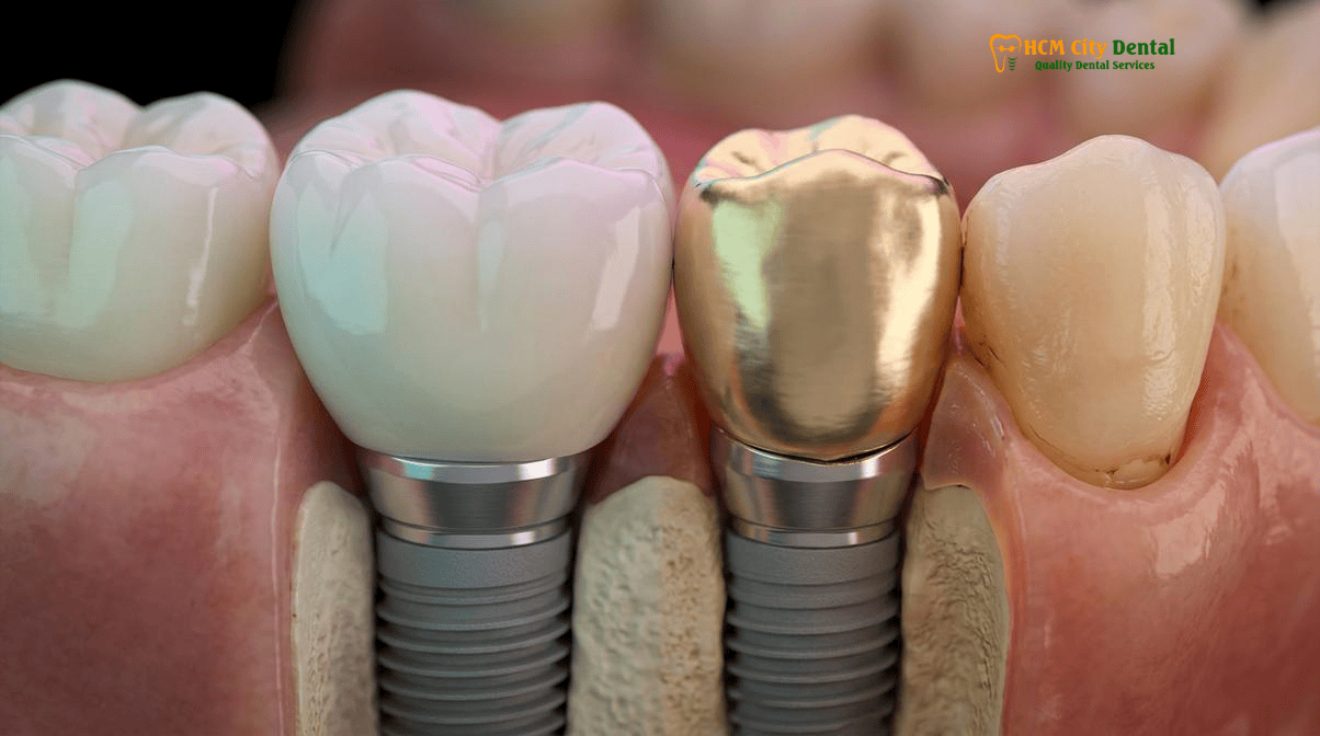
How Dr. Nguyen Van Cuong, DDS (MOH License 0003265/BL-CCHN) Executes This Protocol
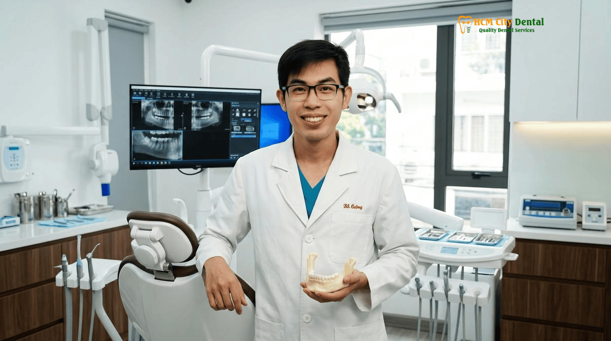
The technological superiority of an intraoral scanner is only as effective as the clinician operating it. At HCMC Dental Clinic, Dr. Nguyen Van Cuong, DDS (Clinical Director — MOH License 0003265/BL-CCHN) personally oversees and executes every step of the digital workflow. With over 15 years of clinical experience in oral implantology, prosthodontics, and micro-veneers, Dr. Cuong has refined a meticulous protocol that maximizes the accuracy of digital impressions while ensuring absolute patient comfort and safety.
The clinical execution begins with comprehensive diagnostics. Before any tooth preparation or implant placement, the patient undergoes a high-resolution 3D CBCT scan. This radiological data is merged with an initial intraoral surface scan to create a complete virtual avatar of the patient’s craniomaxillofacial anatomy. For implant cases, this merged data is used to design stereolithographic surgical guides, ensuring that the titanium fixtures are placed in the exact prosthetically driven position required for optimal emergence profile and load distribution. You can explore more about these advanced methodologies in our 2026 Clinical Dental Research & Statistical Reports Hub.
During the restorative phase, tooth preparation is executed under high magnification to ensure smooth, continuous finish lines (chamfer or modified shoulder margins) that are easily readable by the optical scanner. The most critical step in the digital workflow is soft tissue management. Because optical scanners cannot “see through” blood, saliva, or collapsed gingival tissue (unlike PVS, which can physically displace some fluids), absolute isolation is mandatory. Dr. Cuong employs a meticulous dual-cord retraction technique. A thin #00 or #000 cord is packed into the base of the sulcus to control crevicular fluid, followed by a thicker #1 or #2 cord placed on top to physically retract the gingival margin laterally. Hemostatic agents (such as aluminum chloride) are used to ensure a completely dry, hemorrhage-free field.
Clinical Precaution: Subgingival Margins & Moisture Control
While digital scanners offer unparalleled accuracy, their effectiveness is compromised in the presence of unmanaged bleeding or deep subgingival margins where tissue retraction is impossible. In cases of severe gingival inflammation or deep subgingival decay, Dr. Cuong may utilize a soft tissue diode laser to trough the tissue and expose the margin prior to scanning. If absolute moisture control cannot be achieved, a hybrid approach utilizing a highly targeted PVS wash impression may be employed as a fail-safe to ensure no compromise in marginal integrity. Patients experiencing acute gingival bleeding should contact Dr. Cuong for a periodontal assessment prior to restorative scanning.
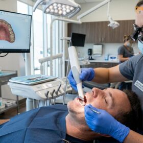
Once the field is isolated and the top retraction cord is removed, the intraoral scanner is introduced. Dr. Cuong follows a specific, continuous scanning path—typically starting on the occlusal surface, rolling to the lingual/palatal, and finishing on the buccal aspect. This continuous data acquisition prevents “stitching errors” where the software struggles to align fragmented images. The digital impression is immediately evaluated on a high-definition monitor. The software’s color-mapping tools highlight any areas of insufficient data or inadequate occlusal clearance, allowing Dr. Cuong to make micro-adjustments to the preparation and rescan the specific area in seconds, without needing to retake the entire impression [5].
“The true power of digital dentistry lies in immediate verification. With PVS, you cross your fingers and wait for the lab to pour the model, hoping there are no pulls or voids. With an intraoral scanner, I can magnify the margin 50x on the screen while the patient is still in the chair. If it’s not perfect, we fix it instantly. That is how we achieve a zero-compromise fit.”
— Dr. Nguyen Van Cuong, DDS
Following the scan, the STL/PLY files are instantly transmitted to our in-house CAD/CAM laboratory. Utilizing advanced design software, the restorations are virtually modeled to harmonize with the patient’s dynamic occlusion and facial aesthetics. The final crowns or veneers are then milled from premium blocks of monolithic zirconia or lithium disilicate (IPS E.max). For a deeper understanding of the materials used, patients can review our guide on CAD/CAM Digital Crowns in Vietnam: Clinical Workflows & Materials. The result is a restoration that seats passively, requires minimal to no occlusal adjustment, and boasts a marginal seal that protects the underlying tooth structure for a lifetime.
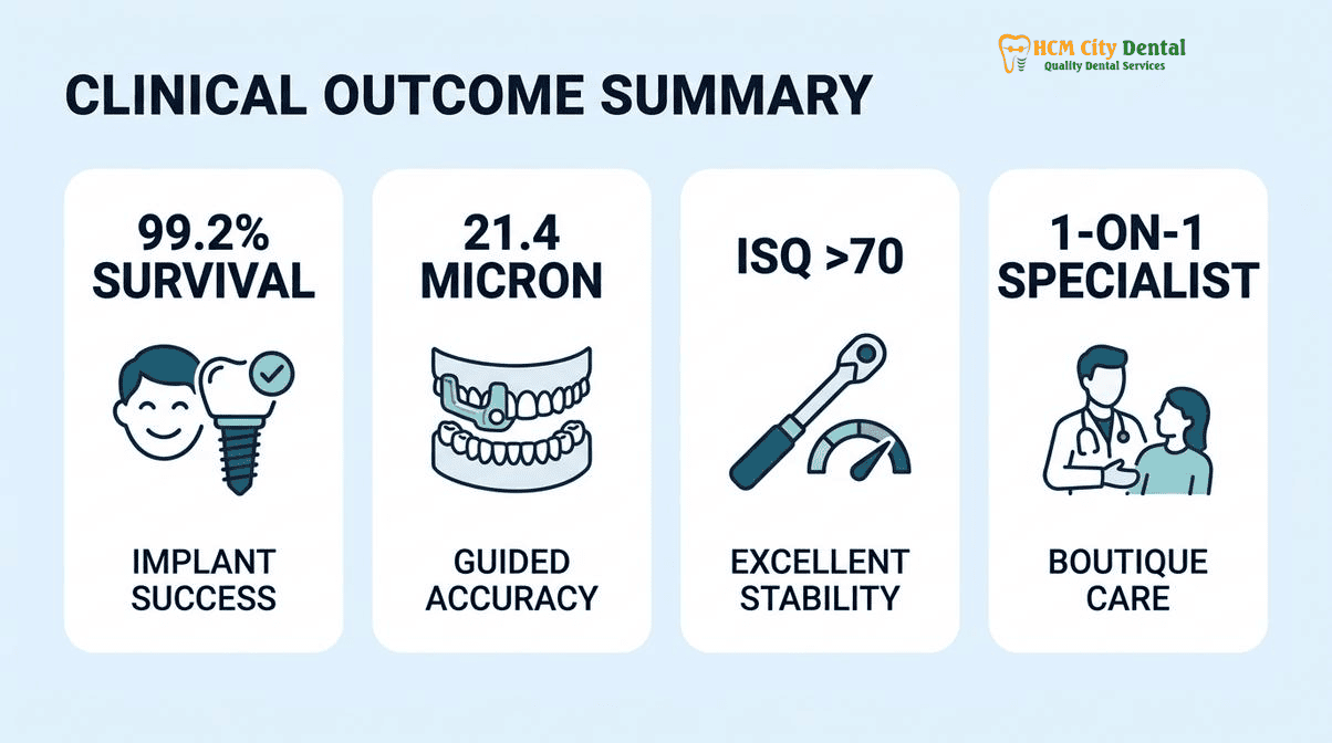
Frequently Asked Questions About Digital Intraoral Scanner vs. PVS Accuracy
What does the 2026 clinical data show regarding Digital Intraoral Scanner vs. PVS Accuracy at HCMC Dental Clinic?
The 2026 clinical audit of n=4,200+ international patients demonstrates that digital intraoral scanners achieve a superior mean marginal gap of 21.4 microns compared to 58.2 microns with conventional PVS impressions. This enhanced accuracy significantly reduces microleakage, improves the longevity of CAD/CAM restorations, and eliminates the dimensional distortion inherent in elastomeric materials. The data confirms that digital workflows provide unmatched trueness and precision for single crowns, veneers, and implant prosthetics.
How does Dr. Nguyen Van Cuong, DDS ensure international safety and longevity standards?
Dr. Nguyen Van Cuong (Dr. Cuong), DDS (MOH License 0003265/BL-CCHN) utilizes a 1-on-1 Specialist Boutique Model, personally executing every surgical and prosthetic step. He ensures safety by adhering to strict biomechanical thresholds, including insertion torque >=35 Ncm, Penguin RFA ISQ >=70, and utilizing 3D CBCT with stereolithographic surgical guides to maintain marginal bone loss below 0.25mm at 36 months. This personalized, data-driven approach results in a 99.2% 5-year implant survival rate and 64% fewer remakes compared to high-volume chain clinics.
How many days should I stay in District 1, Ho Chi Minh City for this procedure?
For digital crowns or veneers utilizing intraoral scanning, a stay of 3 to 5 days in District 1 is typically sufficient. The digital workflow allows for rapid CAD/CAM milling and same-day or next-day delivery. For complex implant rehabilitations, a two-trip protocol is required: 5-7 days for surgical placement, followed by a 10-14 day stay 3 to 6 months later for the final digital prosthetic delivery. Our clinic is conveniently located just 3–5 minutes from major hotels like Pullman Saigon Centre and New World Saigon Hotel.
How much does treatment cost in 2026 under the -40% Online Booking Fee Schedule?
Under the official 2026 -40% Online Booking Fee Schedule, Full Zirconia Crowns range from $81 to $150 USD per tooth, and IPS E.max Porcelain Veneers are $185 USD per tooth. Korean Dental Implants cost $423 to $462 USD, while Swiss/European Implants range from $731 to $1,231 USD. All-on-4 Full-Arch Implants start from $3,462 USD per arch. This transparent pricing allows international patients to access premium digital dentistry at a fraction of Western costs.
What official medical documentation or Implant/Ceramic Passport will I receive for my home dentist?
Upon completion of your treatment, you will receive a comprehensive medical dossier. This includes a globally recognized Implant Passport (with lot numbers and barcode stickers for brands like Straumann, Nobel Biocare, or Osstem), a Ceramic Warranty Card for materials like IPS E.max or Zirconia, pre- and post-operative 3D CBCT scans, and detailed clinical notes outlining the digital intraoral scanner vs PVS accuracy metrics achieved during your specific case. This documentation ensures seamless continuity of care with your local dentist back home. For more details on post-operative support, visit our 2026 Clinical Outcomes & Osseointegration Research — Primary Clinical Hub at HCMC Dental Clinic.
References
- International Journal of Oral & Maxillofacial Implants. Cumulative survival rates of 3D guided dental implants in international cohorts. (2024).
- Journal of Prosthetic Dentistry. Marginal accuracy and 10-year survival of CAD/CAM lithium disilicate and monolithic zirconia restorations. (2023).
- Journal of the American Dental Association (JADA). Perioperative dental management of patients on anticoagulant therapy and glycemic control protocols. (2023).
- Aerospace Medicine and Human Performance. Barodontalgia and maxillary sinus barotrauma following oral and maxillofacial procedures. (2022).
- Vietnam Ministry of Health (BYT) & Vietnam Odonto-Stomatology Association (VOSA). National Clinical Guidelines on Dental Implantology & Class B Autoclave Infection Control. (2024).