Frenular restrictions—commonly known as tongue-ties (ankyloglossia) and lip-ties—are congenital anatomical anomalies where the mucosal attachments in the oral cavity are excessively short, thick, or tight. Historically dismissed as minor pediatric issues that children would simply “outgrow,” modern dental medicine now recognizes that untreated tethered oral tissues (TOTs) trigger a cascade of lifelong functional deficits. In infants, a restricted lingual frenum severely impairs breastfeeding mechanics, leading to poor latch, maternal nipple trauma, and failure to thrive. In older children and adults, the consequences of a restricted tongue or upper lip extend far beyond speech impediments. Chronic ankyloglossia forces a low resting tongue posture, which fundamentally alters maxillofacial growth, narrows the dental arches, induces chronic mouth breathing, and significantly increases the risk of obstructive sleep apnea (OSA) and temporomandibular joint (TMJ) myofascial pain. Today, performing a diode laser frenectomy for tongue-tie (ankyloglossia) and lip-tie release in adults and kids has become the gold standard of care. By utilizing advanced 940/980 nm diode or Er:YAG lasers, specialist dentists can vaporize restrictive tissues with micron-level precision, achieving instant hemostasis, zero need for sutures, and profound functional rehabilitation when combined with targeted orofacial myofunctional therapy (OMT).
Clinical Summary:
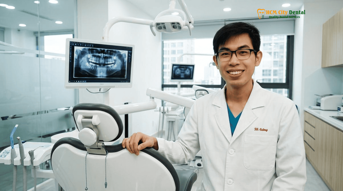
A laser frenectomy is a minimally invasive, bloodless surgical procedure utilizing focused light energy (Diode or Er:YAG lasers) to release restrictive frenular attachments under the tongue (ankyloglossia) or inside the upper lip. At HCMC Dental Clinic in District 1, Ho Chi Minh City, Dr. Nguyen Van Cuong, DDS, employs a comprehensive functional frenuloplasty protocol. This approach not only vaporizes the tethered tissue without scalpels or stitches but also integrates mandatory pre- and post-operative myofunctional therapy. This dual-phase protocol ensures optimal palatal tongue posture, prevents scar reattachment, aids in orthodontic diastema closure, and resolves chronic airway and myofascial tension in both pediatric and adult patients.
Key Takeaways:
- Bloodless Precision: 940/980 nm diode lasers instantly coagulate capillaries, eliminating bleeding, sutures, and severe postoperative pain, making the procedure highly tolerable for anxious children.
- Comprehensive Diagnosis: Accurate assessment requires evaluating both anatomical insertion (Kotlow and Placek classifications) and functional mobility (Tongue Range-of-Motion Ratio).
- Adult Systemic Relief: Releasing an adult tongue-tie dramatically improves palatal resting posture, which opens the posterior airway, reduces snoring, and alleviates chronic cervical and TMJ tension.
- Orthodontic Synergy: Upper labial frenectomies are critical for preventing the relapse of a midline diastema (front tooth gap) after braces or Invisalign treatment.
- Mandatory Aftercare: 21 days of active diamond-wound stretching and myofunctional therapy are clinically required to prevent the rapid reattachment of the oral mucosa.
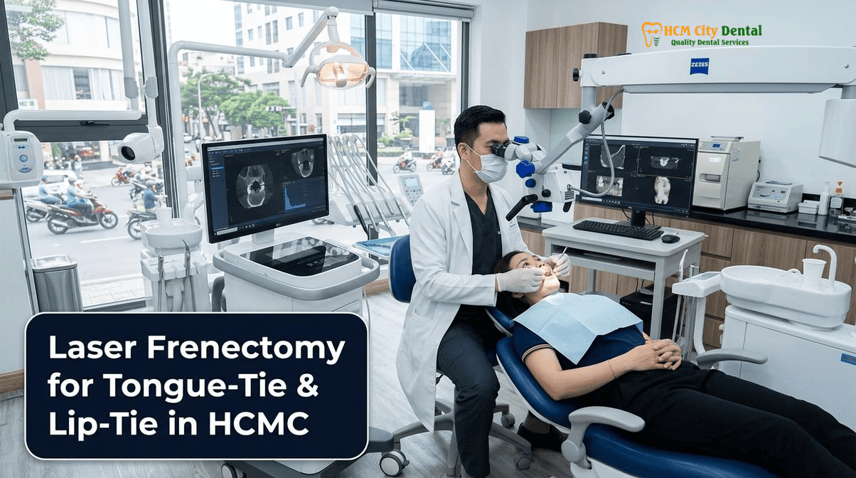
Kotlow’s Anatomical Classification (Class I–IV) & Functional Tongue Range-of-Motion Ratio (TRMR): Diagnosing Anterior vs. Posterior Submucosal Tongue-Tie
The clinical diagnosis of ankyloglossia extends far beyond simply looking under the tongue. A highly restrictive tongue-tie may be visually obvious, attaching directly to the tip of the tongue, or it may be entirely hidden beneath the mucosal surface, tethering the deep genioglossus muscle. To standardize diagnosis, dental specialists utilize Kotlow’s Anatomical Classification, which categorizes the severity of the restriction based on the length of the “free tongue” (the distance from the insertion of the frenum to the tip of the tongue) [1].
- Class I (Mild Ankyloglossia): The free tongue measures 12 to 16 mm. The restriction is minimal but may still cause subtle functional issues during complex speech articulation or swallowing.
- Class II (Moderate Ankyloglossia): The free tongue measures 8 to 11 mm. The frenum attaches further up the ventral surface of the tongue, often causing a noticeable restriction in elevation and protrusion.
- Class III (Severe Ankyloglossia): The free tongue measures 3 to 7 mm. The frenum attaches near the tip of the tongue, frequently causing a classic “heart-shaped” or V-shaped cleft at the tongue tip upon protrusion.
- Class IV (Complete Ankyloglossia): The free tongue is less than 3 mm. The tongue is essentially fused to the floor of the mouth, severely debilitating feeding, speech, and airway development.
However, anatomical appearance alone is insufficient. A patient may have a visually normal frenum but suffer from a “posterior submucosal tongue-tie,” where dense fascial bands restrict movement beneath the surface. To evaluate this, clinicians measure the Functional Tongue Range-of-Motion Ratio (TRMR). This is calculated by measuring the maximum incisal opening (MIO) and comparing it to the maximum opening achievable while the tongue tip is held against the incisive papilla (the roof of the mouth). A TRMR of less than 50% indicates a severe functional restriction, regardless of the Kotlow classification. Accurate diagnosis using both anatomical and functional metrics is the cornerstone of successful Best Dental Clinics & Specialist Comparison at HCMC Dental Clinic protocols, ensuring that the laser release targets the exact fascial restrictions hindering mobility.
Upper Labial Frenum & Midline Diastema: Identifying a Low Papilla-Penetrating Frenum (Placek Class III/IV) and Positive Blanching Test
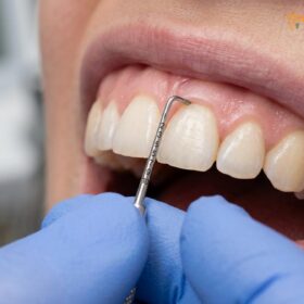
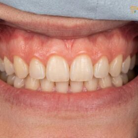
While the lingual frenum dictates tongue mobility, the maxillary labial frenum—the band of tissue connecting the upper lip to the gums—plays a critical role in anterior dental aesthetics and periodontal health. A hypertrophic (enlarged) or low-attaching labial frenum is the primary anatomical culprit behind a persistent midline diastema (a gap between the two upper central incisors). To evaluate the severity of a lip-tie, clinicians utilize the Placek Classification system [2]:
- Class I (Mucosal): The frenum attaches high up in the mucogingival junction, posing no threat to the teeth or gums.
- Class II (Gingival): The frenum inserts into the attached gingiva but remains above the interdental papilla.
- Class III (Papillary): The frenum extends directly into the interdental papilla between the central incisors, often causing the papilla to become inflamed or recede.
- Class IV (Papilla Penetrating): The frenum crosses over the alveolar ridge and inserts directly into the palatal papilla. This severe attachment acts as a thick, fibrous wedge that physically forces the central incisors apart.
To confirm whether a Class III or IV frenum is actively causing a diastema or threatening periodontal health, the clinician performs a “Blanching Test” (also known as the Ischemia Test). By grasping the upper lip and pulling it upward and outward, the dentist observes the interdental papilla and palatal tissues. If the tissue between or behind the front teeth turns white (blanches), it confirms that the frenular fibers are crossing the alveolar ridge and exerting pathological tension on the periodontium. In orthodontic scenarios, performing an upper labial frenectomy is mandatory to prevent the relapse of a closed diastema. However, timing is critical: the laser release must be performed after the orthodontic closure of the gap to prevent scar tissue from impeding tooth movement. For patients seeking comprehensive aesthetic rehabilitation, this procedure is often combined with advanced gingival reshaping by a Laser Gum Contouring & Gummy Smile Correction Specialist Vietnam.
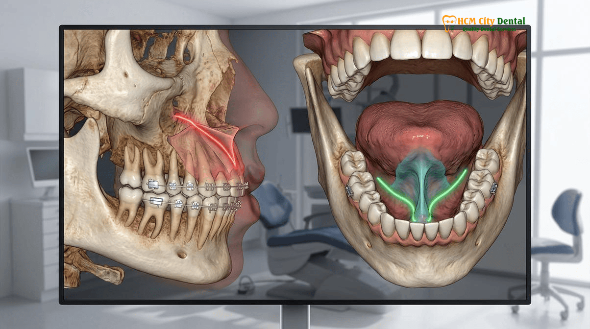
Adult Ankyloglossia, Mouth Breathing & TMJ Myofascial Tension: Why Restricted Lingual Palatal Posture Contributes to Snoring and Cervical Strain
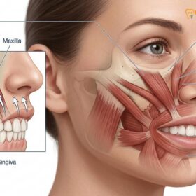
Ankyloglossia is frequently mischaracterized as an exclusively pediatric condition. In reality, thousands of adults suffer from undiagnosed tongue-ties that profoundly impact their systemic health, sleep quality, and musculoskeletal comfort. The human tongue is not merely a muscle for tasting and speaking; it is the structural scaffolding of the maxilla and the upper airway. In a healthy adult, the resting posture of the tongue should be fully suctioned against the hard palate, with the lips sealed and breathing occurring exclusively through the nose.
When an adult has a restricted lingual frenum, the tongue is physically tethered to the floor of the mouth. This low resting posture initiates a devastating domino effect [3]. Without the outward pressure of the tongue, the maxillary arch often develops a narrow, high-vaulted V-shape. To accommodate the low tongue, the mandible drops open, forcing the patient into chronic mouth breathing. Mouth breathing bypasses the nasal filtration system, dries out the oral microbiome (increasing caries risk), and most critically, allows the base of the tongue to collapse backward into the pharynx during sleep. This posterior collapse narrows the airway, drastically increasing airway resistance, turbulent AirFlow (snoring), and the frequency of apneic events (Obstructive Sleep Apnea).
Clinical Case Study: Adult Airway & TMJ Rehabilitation
Patient Profile: A 34-year-old Australian expat presented to HCMC Dental Clinic in District 1 complaining of chronic TMJ pain, severe neck tension, and loud snoring that disrupted his sleep. He had previously tried occlusal night guards with minimal relief.
Clinical Assessment: Dr. Nguyen Van Cuong, DDS, conducted a comprehensive functional exam. The patient exhibited a Class III Kotlow tongue-tie with a TRMR of only 38%. The deep front fascial line was chronically strained, pulling the hyoid bone downward and causing compensatory tension in the sternocleidomastoid and suboccipital muscles.
Treatment Protocol: The patient underwent a 15-minute Er:YAG laser functional frenuloplasty, preceded by four weeks of myofunctional therapy to strengthen the genioglossus muscle.
Outcome: Within 6 weeks post-release, the patient achieved full palatal tongue posture. The chronic TMJ clicking and cervical neck strain resolved entirely, and his sleep partner reported a 90% reduction in snoring volume, highlighting the profound systemic impact of releasing tethered oral tissues.
Furthermore, the tongue is anatomically connected to the “Deep Front Line” of fascia, which runs from the mandible down through the hyoid bone, the pericardium, and into the diaphragm. A tight lingual frenum creates chronic downward tension on the hyoid bone. To keep the airway open and the eyes level, the body compensates by engaging the cervical neck muscles and the muscles of mastication (masseter and temporalis). This compensatory mechanism is a primary, yet frequently overlooked, driver of chronic TMJ disorders, tension headaches, and forward head posture. Releasing the tie via laser surgery, followed by targeted Lip Exercises for Gummy Smile: Clinical Guide & Myofunctional Therapy, restores proper biomechanics, widens the airway, and relieves decades of accumulated myofascial strain.
940/980 nm Diode & Er:YAG Laser Frenectomy vs. Conventional Scalpel & Scissors: Instant Coagulation, Bactericidal Sealing, and Zero Sutures for Children
The surgical approach to releasing tethered oral tissues has undergone a paradigm shift. Historically, a frenectomy was performed using a conventional steel scalpel or surgical scissors. This archaic method severed highly vascularized mucosal tissues, resulting in profuse bleeding that obscured the surgical field. To achieve hemostasis, the surgeon was forced to place multiple sutures (stitches) in the delicate floor of the mouth or under the lip. For pediatric patients, the sight of needles, the taste of blood, and the discomfort of pulling stitches created severe psychological trauma and dental anxiety. Furthermore, scalpel incisions carry a higher risk of postoperative infection, significant swelling, and the formation of thick, restrictive scar tissue.
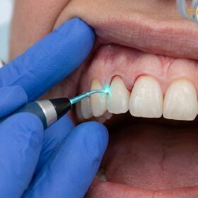
Today, the integration of 940/980 nm soft-tissue diode lasers and Er:YAG all-tissue lasers has revolutionized the frenectomy protocol [4]. Diode lasers operate at wavelengths that are highly absorbed by melanin and hemoglobin. As the laser fiber gently contacts the frenum, the photothermal energy instantly vaporizes the restrictive collagen fibers while simultaneously coagulating the surrounding capillaries and sealing lymphatic vessels. This results in a completely bloodless surgical field, allowing the specialist to visualize and release the deepest fascial restrictions with micron-level precision.
“The transition from scalpel surgery to diode and Er:YAG laser technology is not merely an upgrade in instrumentation; it is a fundamental transformation of the patient experience. By achieving instant photocoagulation and bactericidal sealing, we eliminate the need for sutures and drastically reduce the inflammatory cascade. For an anxious child or a mother struggling with breastfeeding, a bloodless, 5-minute laser release is nothing short of life-changing.”
— Dr. Nguyen Van Cuong, DDS (Clinical Director, HCMC Dental Clinic)
Beyond hemostasis, laser energy provides profound biological advantages. The intense light energy sterilizes the surgical site, destroying pathogenic bacteria and virtually eliminating the risk of postoperative infection. Additionally, the phenomenon of photobiomodulation (low-level laser therapy effects at the periphery of the surgical site) stimulates mitochondrial ATP production, accelerating cellular turnover and reducing postoperative pain signaling. Because no sutures are required, the tissues are allowed to heal via secondary intention, which, when managed correctly with active stretching, results in highly elastic, functional mucosa rather than rigid scar tissue.
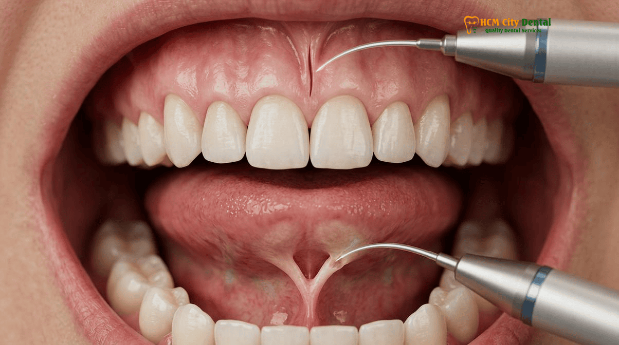
Why Pre- and Post-Operative Orofacial Myofunctional Therapy (OMT) & Diamond-Wound Active Stretches Are Mandatory to Prevent Scar Reattachment
A critical misconception in the treatment of ankyloglossia is that the laser surgery itself is the “cure.” In reality, the laser release is merely the surgical unlocking of the door; Orofacial Myofunctional Therapy (OMT) is the process of walking through it. The oral mucosa is one of the fastest-healing tissues in the human body. When a restrictive frenum is vaporized with a laser, it leaves behind a classic “diamond-shaped” wound. If left alone, the body’s natural inflammatory response will deploy fibroblasts to rapidly bridge this gap, causing the edges of the diamond to contract and heal back together. This phenomenon, known as reattachment, often results in a fibrotic scar that is tighter and more restrictive than the original congenital tie.
To prevent reattachment and ensure the wound heals open and flat, a strict protocol of active stretching must be initiated immediately post-surgery and continued for a minimum of 21 days. For infants, parents are trained to perform gentle but firm lifting and sweeping motions under the tongue and lip every 4 to 6 hours. For older children and adults, active myofunctional exercises are prescribed. These include the “Tongue to Spot” hold, the “Cave” stretch (suctioning the entire tongue to the palate and opening the jaw wide), and lateralization exercises.
Clinical Precaution: The Importance of Compliance
Failure to perform the prescribed post-operative stretches is the single most common cause of frenectomy failure and the need for revision surgery. The stretches may cause mild discomfort during the first 48 to 72 hours, but they are absolutely non-negotiable. If you observe the diamond-shaped wound shrinking rapidly, turning thick and white (beyond normal fibrin slough), or if tongue mobility begins to regress, contact Dr. Nguyen Van Cuong, DDS immediately for clinical evaluation and potential manual disruption of the early adhesions.
Furthermore, pre-operative OMT is highly recommended for adult patients. A tongue that has been tethered for decades suffers from severe muscular atrophy and poor neuromuscular coordination. Engaging in 4 to 6 weeks of myofunctional therapy prior to the laser release strengthens the genioglossus and intrinsic tongue muscles, ensuring that once the physical restriction is removed, the tongue has the strength and muscle memory required to immediately adopt and maintain a healthy palatal resting posture.
Objective 4-Model Clinical Comparison: Choosing the Right Dental Clinic in Ho Chi Minh City for Laser Surgery
For international expats, digital nomads, and dental tourists seeking a precision laser frenectomy in Vietnam, navigating the healthcare landscape can be overwhelming. Ho Chi Minh City features four distinct dental operating models. Understanding these models is crucial for ensuring surgical safety, continuity of care, and transparent pricing [5].
- Model 1: The 1-on-1 Doctor-Direct Specialist Boutique (HCMC Dental Clinic, District 1)
This model is defined by absolute clinical continuity. At HCMC Dental Clinic, every phase of the procedure—from the initial 3D CBCT airway assessment and TRMR measurement to the diode laser surgery and post-op myofunctional follow-up—is performed personally by Dr. Nguyen Van Cuong, DDS (Clinical Director — MOH License 0003265/BL-CCHN). Operating out of a state-of-the-art facility in District 1, this model eliminates associate hand-offs, ensures strict adherence to international sterilization protocols, and offers direct online booking that bypasses agency commissions, resulting in transparent, -40% reduced fees. - Model 2: Multi-Doctor Expat Clinics & Private Dental Hospitals
Facilities such as Westcoast International, Starlight, Elite Dental, and Worldwide Hospital operate primarily in District 1, District 3, and the Thao Dien expat enclave. These clinics offer excellent, Western-standard care and luxurious amenities. However, their massive corporate overhead, extensive marketing budgets, and multi-tiered staffing structures dictate premium pricing. Patients may also experience doctor rotation, where the specialist who performs the consultation is not the same surgeon who performs the laser release or the follow-up care. - Model 3: High-Volume Domestic Dental Chains
Large domestic networks like Kim Dental, Peace Dentistry, and Nhan Tam Dental Clinic operate dozens of branches across Vietnam. While highly accessible and budget-friendly for routine domestic care, these high-volume chains are optimized for general dentistry (fillings, basic extractions). They often lack the specialized 940/980 nm diode or Er:YAG laser technology and the dedicated myofunctional therapy protocols required for successful, non-relapsing functional frenuloplasties. - Model 4: Third-Party Medical Tourism Booking Agencies
Platforms such as Dental Departures or WhatClinic are not healthcare providers; they are referral brokers. They route international patients to partner clinics in exchange for a 15% to 25% commission. Booking through an agency inflates the total cost of treatment and complicates medical liability. For specialized surgical procedures, booking directly with a Ministry of Health (MOH)-licensed clinic ensures direct warranty accountability and eliminates hidden broker fees.
For a deeper analysis of how to select the optimal provider for your specific surgical and aesthetic needs, review our comprehensive Best Dentist & Dental Clinics in Ho Chi Minh City (2026 Comparison Guide).
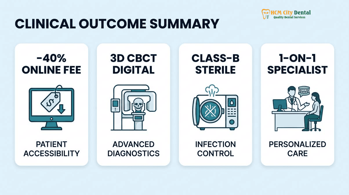
2026 Laser Lingual & Labial Frenectomy Fee Schedule for Children & Adults at HCMC Dental Clinic (District 1)
At HCMC Dental Clinic, we believe that life-changing functional laser surgery should be accessible and transparently priced. By operating a highly efficient, doctor-direct boutique model in District 1, we eliminate corporate bloat and third-party agency commissions. This allows us to offer our international patients an exclusive -40% Online Booking Pricing tier. Below is our official 2026 fee schedule, demonstrating the exceptional value of receiving world-class laser dentistry in Vietnam compared to the exorbitant costs in the US, Australia, or Europe.
| Procedure / Treatment Type | 2026 Online Booking Price (USD) | 2026 Online Booking Price (VND) | Standard Walk-In / US Benchmark |
|---|---|---|---|
| Diode Laser Frenectomy (Lingual / Tongue-Tie) | $58 – $115 USD | 1,500,000 – 3,000,000 VND | $150 Walk-in / $600+ US |
| Diode Laser Frenectomy (Labial / Lip-Tie) | $58 – $115 USD | 1,500,000 – 3,000,000 VND | $150 Walk-in / $500+ US |
| Er:YAG Advanced Laser Frenuloplasty (Complex Adult) | $115 – $154 USD | 3,000,000 – 4,000,000 VND | $250 Walk-in / $800+ US |
| 3D CBCT Airway & Maxillofacial Scan | $18 USD | 480,000 VND | $30 Walk-in / $250+ US |
| Ultrasonic Scaling & Polishing (Pre-Surgical Prep) | $15 USD | 400,200 VND | $25 Walk-in / $150+ US |
| Swiss EMS AirFlow Stain Removal (Erythritol GBT) | $58 USD | 1,500,000 VND | $96 Walk-in / $250+ US |
| Invisalign Comprehensive (Post-Frenectomy Diastema Closure) | $2,538 USD | 66,000,000 VND | $4,231 Walk-in / $6,500+ US |
| Invisalign Vivera Retainer (Genuine SmartTrack) | $308 USD | 8,000,000 VND | $513 Walk-in / $800+ US |
“Transparency in medical billing is a core tenet of our clinical philosophy. By publishing our exact 2026 fee schedule online, we empower our international patients to make informed healthcare decisions without the fear of hidden costs or bait-and-switch tactics. Our -40% online booking rate ensures that premium, bloodless laser surgery remains accessible to everyone.”
— Dr. Nguyen Van Cuong, DDS
To secure these exclusive rates and schedule your comprehensive airway and frenum evaluation, please visit our Official 2026 Dental Fee Schedule (-40% Online Booking Pricing) page or contact our international coordination team directly via WhatsApp at +84 853 020 003.
Frequently Asked Questions
Why does laser frenectomy cause virtually zero bleeding and eliminate the need for stitches in anxious children?
Unlike traditional scalpels that sever blood vessels and require sutures for hemostasis, 940/980 nm diode lasers and Er:YAG lasers utilize photothermal energy to instantly vaporize the restrictive frenum tissue while simultaneously coagulating capillaries. This photocoagulation creates a bloodless surgical field, eliminating the need for terrifying needles or stitches, which significantly reduces intraoperative anxiety and postoperative discomfort for pediatric patients.
When closing a front tooth gap (midline diastema) with braces or Invisalign, should the upper lip-tie laser frenectomy be done before or after space closure?
In modern orthodontic protocols, an upper labial frenectomy is typically performed after the midline diastema has been fully closed with braces or clear aligners. If the frenectomy is performed before space closure, the resulting scar tissue can become dense and fibrotic, making it mechanically difficult to move the central incisors together. Releasing the frenum after closure ensures the tissue heals in its new, tension-free position, preventing orthodontic relapse.
How can releasing a restricted adult tongue-tie improve palatal tongue posture, neck tension, and sleep breathing quality?
A restricted lingual frenum tethers the tongue to the floor of the mouth, preventing it from resting against the hard palate. This low tongue posture forces the mandible to drop, encouraging mouth breathing and narrowing the posterior airway space, which exacerbates snoring and obstructive sleep apnea. Furthermore, because the tongue is anatomically connected to the deep front fascial line, chronic restriction causes compensatory tension in the suprahyoid and cervical muscles. Releasing the tie allows proper palatal posture, widening the airway and relieving chronic neck and TMJ strain.
Why are 21 days of gentle post-laser tongue-lifting exercises critical to prevent the diamond-shaped wound from healing back together?
The oral mucosa heals incredibly fast via secondary intention. After a laser frenectomy, a diamond-shaped wound is left under the tongue or lip. If the tissues are not actively stretched, fibroblasts will rapidly bridge the gap, causing the wound edges to contract and reattach, often resulting in a scar that is tighter than the original frenum. Performing active myofunctional stretches for 21 days ensures the wound heals open and flat, preserving the newly gained range of motion.
How much does a 15-minute Diode or Er:YAG laser frenectomy cost at HCMC Dental Clinic in District 1?
At HCMC Dental Clinic in District 1, Ho Chi Minh City, a precision diode or Er:YAG laser frenectomy is highly affordable, especially when utilizing our direct online booking system. While exact costs depend on the severity of the tie (Class I-IV) and whether myofunctional therapy is included, our transparent 2026 fee schedule offers up to -40% savings compared to walk-in rates, ensuring international expats and dental travelers receive world-class, bloodless laser surgery at a fraction of Western prices.
References
- Kotlow LA. Ankyloglossia (tongue-tie): a diagnostic and treatment quandary. Quintessence International. (1999).
- Placek M, Skach M, Mrklas L. Significance of the labial frenum attachment in periodontal disease in man. Journal of Periodontology. (1974).
- Zaghi S, Valcu-Pinkerton S, Jabara M, et al. Lingual frenuloplasty with myofunctional therapy: Exploring safety and efficacy in 348 cases. Laryngoscope Investigative Otolaryngology. (2019).
- Olivi G, Signore A, Olivi M, Genovese MD. Lingual frenectomy: comparison between the conventional surgical and laser procedure. Pediatric Dentistry. (2012).
- Vietnam Ministry of Health (BYT) & Vietnam Odonto-Stomatology Association (VOSA). National Clinical Guidelines on Specialist Dental Care, Biomaterial Traceability & EN 13060 Class B Infection Control. (2024).